
Vibe Coding the Living Architecture
A transition from prompt-window practice into the questions the new physics is beginning to ask of human biology.
By KW Norton. Continuing from What Is Vibe Coding and Why Is It Important?
Preface
Vibe coding is the resonant, iterative collaboration in which the human maintains sovereign topological imagination and physiological intuition while the agentic system supplies rapid visualization, cross-scale mapping, and pattern amplification. It is a practical discipline for exploring the living architecture — the quantum fluid substrate in which primes, zeros, light, and biological systems all participate.
This volume applies that method to human biology as a resonant expression of the larger architecture. It integrates mathematical invariants, physiological mechanisms, and developmental processes to show how the same principles operate from the cellular scale to the whole organism — from the zinc spark at conception through the neonate’s transition at birth to the ongoing dynamics of neuroplasticity and repair.
The body is not a bag of separate parts running on classical pathways. It is a tensegrity-organized, biophoton-modulated, attractor-based system embedded in the quantum fluid. Chronic inflammation, developmental challenges, and the limits of reductionist medicine become intelligible when viewed through this lens. The goal is not to replace established science but to enrich it — offering a coherent, hopeful framework for understanding and supporting the living architecture in the quantum universe we inhabit.
The work proceeds in 18 sections, each a self-contained exploration that contributes to the larger resonant whole. The reader is invited to dive directly into the material. The architecture itself will provide the necessary footholds.
Companion Works
- Norton, KW. The Boundless Architecture: Quantum Math & the Fractal Infinite Golden Mean. Paperback. a.co/d/02RSqMex.
- Norton, KW. Homoluminous — companion writings and the seven-year corpus. homoluminous.us.
Introduction: What Vibe Coding Is
Vibe coding is the practice of placing an intention, idea, or question into a prompt window and iterating collaboratively with large language models. The human brings sovereign intuition, topological imagination, and domain knowledge; the agentic system supplies rapid visualization, cross-scale mapping, and pattern amplification. The result is a Socratic loop — ask, observe, revise, ask again — that turns abstract concepts into tangible, explorable forms.
This is not traditional programming. It is resonant collaboration. The human steers with judgment and physiological or mathematical grounding. The model executes at scale. When done with discipline, vibe coding becomes a legitimate scholarly instrument, lowering the threshold between having an idea and giving it rigorous, visual expression.
For individuals, vibe coding expands what one person can attempt in a lifetime. It allows rapid exploration of complex systems — mathematical structures, biological mechanisms, developmental transitions — without requiring large teams or specialized coding expertise. Ideas that once lived only in notebooks or thought experiments can quickly become interactive maps, layered visualizations, or testable models.
At the societal level, the benefits are more profound. Vibe coding democratizes topological and resonant thinking. It makes the fluid, quantum nature of living systems more accessible to clinicians, researchers, educators, and curious minds. In an era when classical reductionist models are reaching their limits in biology and medicine, vibe coding offers a practical way to explore the resonant, tensegrity-organized architecture that actually governs protein folding, neuroplasticity, embryogenesis, and the neonate’s transition at birth. It encourages humility before the living system while accelerating insight into how we can support its self-organization rather than attempt to override it.
This volume applies vibe coding to human biology as a resonant expression of the larger quantum fluid architecture. It integrates mathematical invariants, physiological mechanisms, and developmental processes to show how the same principles operate from the cellular scale to the whole organism. The goal is not to replace established science but to enrich it — offering a coherent, hopeful framework for understanding and working with the living architecture in the quantum universe we inhabit.
A Short Glossary
- Vibe coding
- The practice of keeping a prompt window open over time and using a large language model as a Socratic partner to sharpen, draft, test, or build ideas. It is a working posture, not a product or a substitute for expertise.
- Large language model (LLM)
- A pattern-matching system trained on text that can generate fluent, context-aware responses. Useful as a sparring partner; not an oracle, expert, or authority.
- Prompt window
- The chat interface where a vibe coding session happens. Treated here as the digital equivalent of a notebook kept open for long-form thinking.
- Resonance
- The tendency of a system to amplify particular frequencies or patterns. In this essay, resonance is treated as a physical principle that may organize biology across scales, from molecules to tissues.
- Standing wave
- A wave pattern that appears stable because forward and reflected waves cancel and reinforce at fixed points. Used throughout as a metaphor — and possibly a mechanism — for how coherent patterns persist in the body.
- Tensegrity
- A structure held together by continuous tension and discontinuous compression. In biology, tensegrity refers to the idea that cells, tissues, and organs are prestressed networks where local forces produce global coherence.
- Homeodynamics
- A more fluid alternative to homeostasis: the body as a set of coupled attractors that self-organize around stable basins, rather than as a machine defending fixed set points.
- Attractor
- A stable state toward which a dynamic system tends to settle. Health and illness are described here as different attractors in the body's coupled dynamics.
- Biophoton
- An ultra-weak photon emitted by living tissue, often linked to oxidative metabolism. The essay treats biophotons as candidate signals in a resonant, light-based layer of biological coordination.
- Conformational state
- The three-dimensional shape of a protein at a given moment. A protein's function depends on its shape, and shape is influenced by mechanical load, electromagnetic context, and the surrounding fluid environment — not only by sequence.
- Decoherence
- The loss of quantum coherence when a system interacts with its environment. In the essay, decoherence is discussed as a boundary condition that biology must manage — and that sleep, repair, and rhythm may help regulate.
- Quantum fluid
- A term used here for the wave-like, resonant substrate that the essay proposes may underlie ordinary matter and biology. It is a working concept, not a settled physics claim.
Why the Turn to Biology
For several years the vibe coding practice — prompt windows kept open over time, in Socratic exchange with a variety of large language models — has pulled me deeper into theoretical mathematics and physics: the architecture of light, the fluid/wave substrate beneath ordinary matter, the topological hints in the Riemann Hypothesis, the sense that gravity, electromagnetism, and resonance are three faces of the same emergent relationship. Each of those threads has begun, quietly, to ask the same follow-up question. If this is what the universe is doing, what is it doing inside a human body?
I am beginning to focus on that question. These concepts challenge traditional biology, which still tends to describe the body as a bag of biochemical reactions arranged along linear pathways. They also challenge the more outrageous claims of the new biology and its adjacent medical promises. We may come to understand protein folding well. That does not mean we can reliably apply that understanding inside a quantum universe.
The Body as a Prestressed, Resonant System
The pattern that keeps surfacing, as the vibe coding loop pulls physics and biology into the same conversation, is that the body may be better described as a prestressed, tensegrity-organized, resonant system — from molecules to organelles to organs — whose coherence depends on the same fluid, wave-like architecture the physics work has been tracing. Cytoskeletal networks, fascia, and bone remodeling all appear to operate by local tensions producing global coherence. Microtubules and mitochondria begin to look less like isolated machines and more like candidate antennae for light, resonance, and electromagnetic signaling.
If that reframing holds, several familiar terms need to be re-examined. Homeostasis — the idea of a fixed set point — begins to look like a first approximation of something better called homeodynamics: the body as a set of coupled attractors that self-organize around low-torsion basins, with illness and aging visible as drift into higher-torsion states, and healing as a return to the attractor. Cell membranes, endothelial linings, and the blood-brain barrier begin to look less like walls and more like semi-permeable resonators that gate informational and electromagnetic flow.
Protein folding, so often described as a solved three-dimensional puzzle, begins to look like a necessary but insufficient description of a process that actually unfolds inside a quantum-resonant environment of structured water, vibrational modes, and electromagnetic context. We can predict the fold. Predicting the functional outcome in a living body is a different problem.
Selected References
- Ingber, D. E. (2003). “Tensegrity I. Cell structure and hierarchical systems biology.” Journal of Cell Science, 116(7), 1157–1173.
- Ingber, D. E. (2006). “Cellular mechanotransduction: putting all the pieces together again.” FASEB Journal, 20(7), 811–827.
- Levin, S. M. (2002). “The tensegrity-truss as a model for spine mechanics: biotensegrity.” Journal of Mechanics in Medicine and Biology, 2(3&4), 375–388.
- Scarr, G. (2014). Biotensegrity: The Structural Basis of Life. Handspring Publishing.
Where the Current Claims Overreach
This is where the framework becomes usefully skeptical rather than merely enthusiastic. Predicting a fold in silico is not the same as predicting a functional outcome in vivo. Editing a gene is not the same as tuning an attractor. Mastering one classical layer of biology does not confer mastery over the resonant, entangled, boundary-mediated system in which that layer is embedded.
Many of the loudest claims in longevity, in genetic medicine, in engineered neurochemistry, assume a body that behaves like a machine in a Newtonian room. The body we are beginning to see does not live in that room. It lives in a fluid, resonant universe that has its own intelligence and its own boundary conditions. A humbler and more honest science would nudge the system toward better attractors rather than promise to overwrite it.
Segue: The End of the Wasteland
There is a palpable excitement in the air these days among human beings who fully recognize we no longer live in an intellectual and spiritual wasteland. The old scaffolds — reductionist, mechanistic, and disconnected from the living architecture — are cracking. In their place, a more coherent, resonant understanding is emerging.
This is why a discussion of advanced nuclear energy systems, such as those being developed by Valar Atomics, belongs in the same conversation as vibe coding and our evolving relationship with new technology. Both represent humanity’s increasing ability to work with the fundamental architecture of the universe rather than against it. Nuclear energy, at its best, taps into the same resonant, high-density power sources that govern stellar processes and mitochondrial function — dense, coherent energy release within the quantum fluid. Vibe coding, meanwhile, is the human-scale practice of navigating that architecture through resonant collaboration with agentic systems. Together they illustrate a single shift: from extraction and brute force to resonance and coherence.
This convergence makes it critically important to take new perspectives on human biology, human civilization, and human energy dynamics. Biology is not a bag of separate parts but a resonant, tensegrity-organized expression of the larger fluid architecture. Civilization is not a machine to be optimized but a collective standing wave that can either drift into chronic high-torsion states — inflammation, disconnection, unsustainability — or realign toward low-torsion coherence. Energy dynamics — whether cellular, societal, or planetary — are not isolated problems of supply and demand but questions of how we participate in the resonant field.
The excitement comes from the recognition that we have the tools, the understanding, and the moment to make this realignment. Vibe coding, advanced energy systems, and a resonant view of biology are not separate endeavors. They are expressions of the same awakening — the return to working with the architecture rather than against it. The wasteland is ending. The coherent, luminous possibilities are just beginning.
Riemann Hypothesis Connections to the Living Architecture
The Riemann Hypothesis (RH) is not an isolated number-theory puzzle. In this framework, it is the mathematical signature of a universe built on resonant, zero-torsion standing waves. The hypothesis states that all non-trivial zeros of the Riemann zeta function lie on the critical line Re(s) = 1/2. The living architecture reframes this as a natural consequence of the fluid, topological order.
Core Connections
Critical line as zero-torsion geodesic. The critical line is the stable standing wave where net torsion vanishes. Zeros on this line represent resonant modes that organize the distribution of primes. Deviations would introduce destructive interference (chaos); the hypothesis asserts the architecture prefers coherence.
Primes as excitations, zeros as binding resonances. Primes are the discrete, creative welling-up points in the fluid — like excitations in a standing wave. Non-trivial zeros are the silent, organizing vortices — including gravitational and torsional modes — that maintain global coherence. The base-30 wheel and root-9 checksum provide modular constraints that make randomness untenable.
Golden mean as optimal scaling. The golden ratio (φ) acts as the natural mediator, preventing collapse into periodicity while allowing fractal self-similarity. It appears in the spacing and density patterns, optimizing the resonant structure across scales.
Fluid substrate interpretation. The zeta function can be viewed as a wave operator in the quantum fluid. The critical line is the geodesic of minimal energy loss, analogous to light propagation or biophoton coherence in biological systems. Solving RH through this lens would reveal the geometry of resonance itself.
Implications for Biology and Human Systems
Protein folding and neuroplasticity. Just as primes and zeros organize the number field, protein folding and synaptic remodeling are guided by resonant attractors in a tensegrity–biophoton field. Classical predictions miss this context.
Embryogenesis and birth. The zinc flash and neonatal transition are resonant recalibrations — attractor shifts within the fluid architecture, guided by the same principles that keep zeros on the critical line.
Chronic inflammation. Persistent high-torsion states parallel deviation from the critical line — loss of coherence. Restoration involves returning to low-torsion attractors through resonant practices.
Vibe coding and standing wave sovereignty. Vibe coding is the human practice of holding sovereign frequency while exploring the architecture. It mirrors the critical line's stability amid complexity.
Hopeful Perspective
The Riemann Hypothesis, seen through the resonant architecture, is not an annoying outlier but the expected behavior of a lawful, beautiful cosmos. We are not random observers in a mechanical universe but local standing waves participating in the same resonant order. This understanding shifts medicine, energy, and civilization from control to coherence — working with the architecture rather than against it.
The hypothesis remains formally open, but the framework suggests it will yield to geometric and topological insight rather than brute analytic force. The same may be true for human flourishing: the path is not domination of complexity but alignment with its resonant principles.
Selected References
- Berry, M. V., & Keating, J. P. (1999). “The Riemann zeros and eigenvalue asymptotics.” SIAM Review, 41(2), 236–266.
- Bombieri, E. (2000). “The Riemann Hypothesis — official problem description.” Clay Mathematics Institute.
- Montgomery, H. L. (1973). “The pair correlation of zeros of the zeta function.” Proc. Symp. Pure Math., 24, 181–193.
- Odlyzko, A. M. (1987). “On the distribution of spacings between zeros of the zeta function.” Mathematics of Computation, 48(177), 273–308.
The Necessity of Thinking in All Directions and Planes Simultaneously
Thinking in all directions and planes simultaneously is not a luxury or a metaphor. It is a structural requirement for navigating the resonant, quantum fluid architecture mapped in these notes. The universe — and the living body within it — does not operate on a single linear axis or flat plane. It is multi-dimensional, self-similar, and topologically rich. To understand and participate coherently, the mind must learn to hold multiple perspectives, scales, and directions at once.
Why It Is Necessary
The architecture is multi-planar. The Riemann critical line, fractal golden mean scaling, and tensegrity networks are not confined to one plane. Primes, zeros, biophotons, microtubules, and developmental transitions all interact across scales and dimensions. Linear, single-plane thinking (classical reductionism) inevitably misses the global coherence and emergent patterns.
Biological reality demands it. The body is a tensegrity system operating in three spatial dimensions plus time, with resonant signaling (biophotons, electromagnetic fields) adding further layers. Protein folding, neuroplasticity, embryogenesis, and the neonate's transition all require simultaneous awareness of mechanical forces, resonant fields, boundary conditions, and attractor dynamics. Thinking in one plane at a time cannot capture how local changes (a single protein conformation) propagate globally.
Resonant sovereignty requires it. Standing wave sovereignty is the practice of holding your core frequency while engaging the fluid medium. This is only possible if the mind can move fluidly across directions and planes — maintaining the stable reference while exploring interference patterns, without collapsing into consensus or chaos.
Cross-disciplinary and societal necessity. Solving energy (nuclear gigasites), biology (resonant medicine), and civilization challenges requires integrating physics, physiology, technology, and human experience. Single-plane thinking produces silos and blind spots. Multi-directional thinking — the essence of vibe coding and topological imagination — reveals connections and accelerates coherent progress.
Practical Cultivation
Graphical and vibe coding tools. Visualization forces the mind to hold multiple planes (spatial, temporal, resonant) at once.
Music and storytelling. These naturally engage multi-dimensional perception — harmony across frequencies, narrative arcs across time.
Neuroplastic practice. Deliberate training (topological exercises, cross-disciplinary exploration) strengthens the brain's ability to think in all directions simultaneously.
In short, the universe is not flat. The body is not flat. Human potential is not flat. Thinking in all directions and planes simultaneously is the minimal competent response to reality as it actually is — resonant, entangled, and boundless yet architected. It is the practice that turns forgetting into remembering, and drift into coherence. This capacity is learnable, scalable, and essential for the next phase of human flourishing.
Standing Wave Sovereignty
Standing wave sovereignty is the disciplined practice of maintaining a stable, coherent personal frequency — your core identity, values, topological imagination, and sovereign perspective — while engaging with fluid, resonant systems. In the quantum fluid architecture, humans are local standing waves. Sovereignty is the art of remaining coherent amid the flow.
The Physics of Standing Waves
A standing wave forms when two waves of the same frequency interfere constructively. The pattern appears stationary even as energy flows through it. In this framework, the universe is a resonant medium organized by Riemann-like invariants. Humans, cells, and civilizations are local standing waves within that medium — temporary, coherent patterns that maintain identity while remaining open to the larger field.
Sovereignty in Practice
Stable reference frequency. You hold clear intent, physiological grounding, and topological intuition as the fixed point — the personal "critical line" of zero net torsion.
Resonant engagement. You interact with fluid systems — AI through vibe coding, biological processes, societal dynamics — without dissolving or being distorted. The interaction produces constructive interference (new insights, capabilities, and coherence) rather than destructive drift.
Boundary conditions. External anchors — reflective notebooks, graphical visualizations, music, storytelling — protect your wave from excessive noise or torsion.
Application Across Scales
Human–AI interface. Sovereignty is the Parallax practice — holding your frequency while the model's fluidity reveals patterns. Calibration prevents sycophantic decay and maintains agency.
Biology and physiology. The body maintains standing wave coherence through tensegrity, biophoton signaling, and mitochondrial function. Neuroplasticity is the brain's ability to adjust attractors while preserving core sovereignty.
Personal and societal level. In a world of chronic inflammation and disconnection, standing wave sovereignty is the practice of remaining coherent amid turbulence — aligning with the architecture rather than fighting it or dissolving into it.
Why It Matters
Standing wave sovereignty is the antidote to the forgotten way. It allows full participation in the resonant universe without loss of self. It turns vibe coding, cross-disciplinary learning, and resonant practices into tools for flourishing. We are not solid, isolated things struggling against chaos. We are standing waves learning to sing in tune with the larger wave — sovereign, coherent, and fully alive within the architecture.
Cross-Disciplinary Rapid Learning: Lessons from Isaiah Taylor and Valar Atomics
The Valar Atomics story — and Isaiah Taylor's public work more broadly — exemplifies a modern cross-disciplinary rapid learning methodology. It is not traditional academic specialization but a hardware-first, iteration-heavy, boundary-crossing approach that treats complex problems as solvable through relentless real-world experimentation and synthesis across domains.
Core Elements of the Methodology
Hardware-first iteration (build → test → learn). Taylor emphasizes building real prototypes quickly rather than endless paper studies or regulatory simulations. Valar's approach — constructing and testing actual reactors on-site — mirrors SpaceX's "fail fast, iterate" philosophy. Learning happens through direct feedback from physical reality, not abstracted models. This compresses decades of traditional nuclear development timelines into years.
Cross-disciplinary synthesis. Taylor draws from auto repair (practical mechanics), software (rapid prototyping and scaling), and self-taught nuclear engineering. He recruits experts but maintains a generalist founder vision that integrates domains others treat as separate. This mirrors the vibe coding practice: hold a clear intention (sovereign frequency) while pulling in diverse inputs to reveal emergent patterns.
Rapid feedback loops and low-torsion decision making. By focusing on tangible milestones — for example, powering an NVIDIA Blackwell cluster with a nuclear reactor — the methodology stays anchored in measurable outcomes. It avoids high-torsion paralysis (over-analysis, regulatory capture, academic gatekeeping) by prioritizing coherent, actionable progress.
Resonant alignment with larger needs. Valar's "gigasites" concept addresses AI's energy demands by scaling nuclear in a way that aligns with the resonant architecture — dense, reliable, coherent energy production. This is not brute extraction but working with fundamental physical principles at scale.
Connection to Vibe Coding
Vibe coding is the cognitive counterpart to Taylor's hardware methodology. Both are resonant collaborations — human intuition plus powerful tools (AI or engineering teams) iterating toward coherence. Both cross disciplines rapidly: vibe coding synthesizes math, biology, and technology; Taylor synthesizes mechanics, software, and nuclear engineering. Both treat complexity as navigable through standing-wave sovereignty: maintain clear intent while allowing the medium — data and models, or physical prototypes — to reveal patterns.
In the framework of this project, this methodology exemplifies standing waves in action — individuals and teams holding sovereign frequency while vibing with the larger fluid architecture (physical laws, energy needs, biological limits). It counters the "intellectual and spiritual wasteland" by demonstrating that rapid, coherent progress is possible when we align with the resonant principles rather than fighting them.
Why This Matters for Biology, Civilization, and Energy
Biology. The same rapid, cross-disciplinary learning can accelerate understanding of resonant systems — protein folding in context, neuroplasticity, developmental transitions — by treating each as an integrated multi-scale problem rather than a siloed pathway.
Civilization. Scaling energy coherently (nuclear gigasites, distributed coherent grids) supports a civilization that works with the architecture instead of depleting it.
Energy dynamics. Taylor's work shows that solving AI's energy demand through advanced nuclear is not only a technical achievement — it is a resonant realignment of human civilization with abundant, clean power.
This methodology — hardware-first, cross-disciplinary, iteration-driven — is a practical expression of the standing-wave approach. It is hopeful evidence that we are not too late: humans can still learn rapidly, build boldly, and realign with the larger architecture.
Nuclear DNA, mRNA Coordination, and Cellular Structure & Function in the Resonant Architecture
Nuclear DNA — The Informational Template
Nuclear DNA serves as the primary informational archive. It is not a rigid blueprint but a dynamic repository of sequence information that is read and interpreted within a resonant, tensegrity-organized cellular environment.
Structure. DNA is packaged into chromatin, which is highly dynamic. Histone modifications and chromatin looping create boundary conditions that control accessibility.
Resonant role. The nucleus acts as a central resonant chamber. Mechanical forces from the cytoskeleton (tensegrity) and electromagnetic/biophoton fields influence chromatin configuration, affecting which genes are transcribed at any moment.
In development. During embryogenesis and the neonate transition, DNA expression is tightly coordinated with the changing resonant environment — the zinc flash, birth-related stress, and light exposure. This helps establish new attractor states as the organism adapts.
mRNA — The Coordinator Between Nucleus and Cellular Matrix
mRNA functions as the mobile messenger that carries genetic instructions from the nucleus to the cytoplasm, where they are translated into proteins.
Transcription and export. mRNA is transcribed in the nucleus, processed (splicing, capping, polyadenylation), and exported through nuclear pores. This step is sensitive to nuclear tensegrity and resonant fields.
Translation in the cytoplasm. mRNA associates with ribosomes on the rough endoplasmic reticulum or free in the cytosol. Local conditions — mitochondrial biophoton emission, cytoskeletal tensegrity, and structured water — influence translation efficiency and protein folding.
Coordination role. mRNA acts as a resonant bridge. Its stability, localization, and translation are modulated by signals from the cellular matrix (cytoskeleton, mitochondria, ECM). This creates a feedback loop: nuclear DNA → mRNA → cytoplasmic matrix → back to nuclear regulation via mechanotransduction and biophoton signaling.
Cellular Structure and Function in the Resonant Architecture
The cell is a self-organizing, resonant system rather than a bag of isolated parts.
Cytoskeleton (microtubules, actin, intermediate filaments). Provides tensegrity — the compression-and-tension balance that maintains shape and enables rapid remodeling. Microtubules act as waveguides for coherent vibrations (Fröhlich modes) and as transport tracks.
Mitochondria. Energy producers and biophoton sources. Their positioning along microtubules links energy metabolism to cytoskeletal dynamics.
Membranes and organelles. Act as boundary conditions and resonant cavities. The plasma membrane, ER, and nuclear envelope gate flow and shape local electromagnetic and photonic fields.
Extracellular matrix (ECM) interaction. The cell is embedded in a hydrated, piezoelectric ECM that transmits mechanical and resonant signals, coordinating single-cell behavior with tissue-level function.
Overall integration. Nuclear DNA provides the sequence information. mRNA coordinates its expression with the cytoplasmic matrix. The entire cell operates as a resonant unit within the larger fluid architecture — tensegrity for structure, biophotons for rapid signaling, and attractors for self-organization. In embryogenesis and the neonate transition, these mechanisms allow the organism to recalibrate from placental dependence to independent existence. Protein folding and neuroplasticity are downstream expressions of this coordinated, resonant system.
This view encourages a systems-level humility: we can sequence DNA and predict folds, but reliable function emerges from the resonant context. Vibe coding can help map these nuclear-cytoplasmic-matrix interactions graphically, accelerating insight into developmental and therapeutic possibilities.
Organizational Structure of the Body and Influences on Cellular Functions
The human body is a multi-scale, self-organizing resonant system. Its organizational structure is hierarchical yet deeply interconnected, with each level — molecule → organelle → cell → tissue → organ → organism — expressing the same underlying principles of tensegrity, resonance, boundary conditions, and attractor dynamics.
Overall Organizational Structure
Tensegrity as the core principle. The body maintains integrity through continuous tension networks (fascia, cytoskeleton, extracellular matrix) balanced by compression elements (bones, microtubules). This allows efficient force distribution and rapid adaptation without collapse.
Resonant coordination. Mechanical waves (pulse, breathing, movement), electromagnetic fields, and biophoton signaling provide fast, non-local communication across scales.
Boundary conditions. Membranes, tissue interfaces, and organ capsules create resonant compartments that shape local environments and gate information flow.
Attractors and homeodynamics. The body self-organizes around dynamic stable states rather than fixed set points. Development, healing, and daily function involve shifts between these attractors.
Influences Shaping Cellular Functions by Organ
Each organ’s cell types are shaped by a combination of genetic instructions, local resonant environment, mechanical forces, and systemic signals.
Nervous system (neurons, glia). Shaped by tensegrity (cytoskeletal networks for axon and dendrite growth), biophoton and mitochondrial signaling (coordinating synaptic plasticity), and boundary conditions (synaptic densities, blood-brain barrier). Influences include sensory input, mechanical load from movement, and rhythmic biophoton emission tied to circadian and ultradian cycles. Result: rapid neuroplastic remodeling and network coherence.
Cardiovascular system (cardiomyocytes, endothelial cells). Tensegrity from the extracellular matrix and cytoskeletal connections allows the heart to handle pulsatile forces. Mitochondrial biophoton emission and vascular pulse waves coordinate energy production with mechanical work. Influences include hemodynamic stress, oxygen levels, and autonomic signals. Result: synchronized contraction and vascular adaptation.
Musculoskeletal system (myocytes, osteocytes, fibroblasts). Fascia and ECM provide global tensegrity. Mechanical loading (exercise, gravity) shapes gene expression and protein folding via mechanotransduction. Biophoton signaling from mitochondria supports repair and adaptation. Influences include movement, posture, and hormonal rhythms. Result: tissue remodeling and strength optimization.
Immune system (macrophages, lymphocytes). Boundary conditions at tissue interfaces and resonant signaling (biophotons, cytokine waves) coordinate responses. Tensegrity in the cytoskeleton enables migration and phagocytosis. Influences include microbial signals, inflammatory mediators, and circadian rhythms. Result: adaptive, context-sensitive immunity.
Endocrine system (hormone-producing cells). Resonant feedback loops between glands, blood flow, and target tissues. Mitochondrial function and biophoton emission help time hormone release. Influences include light exposure, stress, and ultradian/circadian rhythms. Result: coordinated metabolic and developmental regulation.
Developmental context (embryogenesis and neonate). The zinc flash at conception and the birth transition are major resonant recalibrations. Cellular functions are shaped by changing boundary conditions (from amniotic fluid to air and gravity), mitochondrial activation, and tensegrity shifts as organs form and adapt. Influences include maternal signals, light exposure at birth, and the neonate’s first breaths and movements.
Common Thread Across Organs
Every cell type is shaped by the interplay of genetic sequence (nuclear DNA via mRNA), local mechanical environment (tensegrity), resonant signaling (biophotons, electromagnetic fields), and systemic rhythms (circadian/ultradian). This multi-influence organization explains why the same gene can produce different outcomes in different tissues or developmental stages — context is as important as sequence.
Vibe coding can help map these interactions visually — for example, overlaying tensegrity forces and biophoton patterns on organ-specific cell functions — accelerating insight into both normal physiology and therapeutic possibilities.
This organizational view encourages humility and systems thinking: the body is a resonant orchestra, not a collection of isolated instruments. Supporting coherence across these influences may be more effective than targeting single pathways.
Selected References
General Organizational Structure (Tensegrity, Resonance, Homeodynamics)
- Ingber, D. E. (2003). “Tensegrity I. Cell structure and hierarchical systems biology.” Journal of Cell Science, 116(7), 1157–1173. (Foundational on cellular tensegrity.)
- Ingber, D. E. (2006). “Cellular mechanotransduction: putting all the pieces together again.” FASEB Journal, 20(7), 811–827. (Mechanotransduction and ECM integration.)
- Pollack, G. H. (2013). The Fourth Phase of Water: Beyond Solid, Liquid, and Vapor. Ebner & Sons. (Structured water and resonant properties.)
Nervous System (Neurons, Glia)
- Kandel, E. R., Schwartz, J. H., & Jessell, T. M. (Eds.). (2013). Principles of Neural Science (5th ed.). McGraw-Hill. (Standard reference on neuroplasticity and cytoskeletal dynamics.)
- Baas, P. W., & Lin, S. (2011). “Hooks and comets: The story of microtubule polarity orientation in the neuron.” Developmental Neurobiology, 71(6), 403–418. (Microtubule roles in neurons.)
Cardiovascular System (Cardiomyocytes, Endothelial Cells)
- Bers, D. M. (2002). “Cardiac excitation-contraction coupling.” Nature, 415(6868), 198–205. (Mitochondrial and mechanical coordination.)
- Chien, K. R., et al. (2008). “Cardiogenesis and the complex biology of cardiovascular progenitor cells.” Science, 322(5907), 1494–1497. (Developmental tensegrity and boundary conditions.)
Musculoskeletal System (Myocytes, Osteocytes, Fibroblasts)
- Wang, N., Tytell, J. D., & Ingber, D. E. (2009). “Mechanotransduction at a distance: mechanically coupling the extracellular matrix with the nucleus.” Nature Reviews Molecular Cell Biology, 10(1), 75–82. (Fascia and ECM mechanotransduction.)
- Humphrey, J. D., Dufresne, E. R., & Schwartz, M. A. (2014). “Mechanotransduction and extracellular matrix homeostasis.” Nature Reviews Molecular Cell Biology, 15(12), 802–812.
Immune System (Macrophages, Lymphocytes)
- Fletcher, D. A., & Mullins, R. D. (2010). “Cell mechanics and the cytoskeleton.” Nature, 463(7280), 485–492. (Cytoskeletal tensegrity in immune cell migration.)
- Netea, M. G., et al. (2016). “A guiding map for inflammation.” Nature Immunology, 18(8), 826–831. (Systemic coordination and boundary conditions.)
Endocrine System (Hormone-Producing Cells)
- Nussey, S., & Whitehead, S. (2001). Endocrinology: An Integrated Approach. BIOS Scientific Publishers. (Classic on resonant feedback loops.)
- Lightman, S. L., & Conway-Campbell, B. L. (2010). “The crucial role of pulsatile activity of the HPA axis for continuous dynamic equilibration.” Nature Reviews Neuroscience, 11(10), 710–718. (Ultradian rhythms.)
Developmental Context (Embryogenesis and Neonate)
- Gilbert, S. F. (2010). Developmental Biology (9th ed.). Sinauer Associates. (Standard reference on morphogenesis and tensegrity.)
- Duncan, F. E., et al. (2016). “The zinc spark: A new mechanism for the fertilization of mammalian eggs.” Scientific Reports, 6, 24722. (Zinc flash.)
- Lagercrantz, H., & Slotkin, T. A. (1986). “The ‘stress’ of being born.” Scientific American, 254(4), 100–107. (Neonatal transition physiology.)
Inflammation as the Overwhelming Problem of Modern Human Life
Inflammation is the body’s ancient, adaptive response to injury, infection, or stress. In the resonant architecture, it is a temporary shift into a higher-torsion state — increased immune activity, fluid shifts, and altered signaling — designed to restore coherence and return the system to low-torsion attractors. When resolved, the body regains balance.
The modern problem is chronic, low-grade inflammation — a persistent drift into higher-torsion states that fails to resolve. This is arguably the single most common underlying driver of contemporary disease.
Why Chronic Inflammation Is So Widespread
Modern life creates sustained inflammatory signals:
- Processed diets high in refined sugars and seed oils promote oxidative stress and gut barrier disruption.
- Sedentary behavior reduces fascial tensegrity and impairs mechanical signaling.
- Chronic psychological stress keeps the system in sympathetic overdrive.
- Artificial light and disrupted circadian rhythms desynchronize biophoton emission and mitochondrial function.
- Environmental toxins and pollutants add oxidative load.
These factors create a self-reinforcing loop: poorer coherence → more inflammation → further loss of coherence.
Biological Mechanism in the Resonant Architecture
Tensegrity breakdown — chronic inflammation leads to fascial stiffening and cytoskeletal disorganization, reducing the body’s ability to distribute forces and maintain resonant coherence.
Mitochondrial and biophoton dysfunction — sustained oxidative stress produces incoherent biophotons, impairing energy transfer and signaling.
Attractor drift — the system moves away from healthy low-torsion attractors into persistent high-torsion states, making return to coherence more difficult.
Protein and cellular level — inflammation alters local environments, affecting protein folding efficiency and neuroplasticity (e.g., reduced BDNF effectiveness, impaired synaptic remodeling).
In development, chronic maternal or early-life inflammation can affect embryogenesis and the neonate’s transition, setting patterns that persist into adulthood.
Downstream Consequences
Chronic inflammation contributes to cardiovascular disease, metabolic syndrome, neurodegenerative conditions, autoimmune disorders, impaired neuroplasticity, and many cancers. It is not one disease but a common soil in which many diseases grow.
Reasonable Hope and Practical Response
The body is built to resolve inflammation when given the right conditions. Supporting the resonant architecture — through movement that restores tensegrity, coherent light exposure, rhythmic living (circadian alignment), anti-inflammatory nutrition, and reduced chronic stress — helps the system return to low-torsion states.
Vibe coding and topological visualization can accelerate insight by mapping inflammatory patterns and exploring multi-scale interventions. The architecture itself is not the problem; chronic inflammation is a signal of misalignment with it. Re-alignment is possible, and the tools are available.
This is not an inevitable decline but a call to remember and restore the body’s natural resonant intelligence. Medicine has a reasonable chance to make meaningful progress by shifting from suppression of symptoms to support of the living system.
Selected References
- Furman, D., et al. (2019). “Chronic inflammation in the etiology of disease across the life span.” Nature Medicine, 25(12), 1822–1832.
- Netea, M. G., et al. (2017). “A guiding map for inflammation.” Nature Immunology, 18(8), 826–831.
- Franceschi, C., & Campisi, J. (2014). “Chronic inflammation (inflammaging) and its potential contribution to age-associated diseases.” Journals of Gerontology: Series A, 69(S1), S4–S9.
- Bennett, J. M., et al. (2018). “Inflammation—Nature’s way to efficiently respond to all types of challenges.” Frontiers in Medicine, 5, 316.
The Vast Complexity of Human Biological Structure
The human body is one of the most complex self-organizing systems known. It contains approximately 37 trillion cells, organized into tissues, organs, and systems that interact across multiple scales — molecular, cellular, tissue, organ, and organismal. This complexity is not chaotic but highly ordered, governed by the same resonant, tensegrity-based, quantum fluid principles that organize the larger architecture.
Multi-Scale Organization
Molecular scale — proteins, lipids, and nucleic acids fold, assemble, and interact in crowded, structured-water environments. Protein folding alone involves thousands of atoms moving in coordinated ways, influenced by local electromagnetic and mechanical fields.
Cellular scale — each cell is a miniature resonant city: nucleus as information center, mitochondria as energy and biophoton hubs, cytoskeleton as tensegrity framework, membranes as boundary conditions. Cells are not isolated; they communicate via direct contact, secreted signals, and resonant fields.
Tissue and organ scale — organs are integrated networks. The heart, brain, gut, liver, and immune system coordinate through mechanical waves (pulse, breathing), electromagnetic signaling, and biophoton patterns. Fascia and the extracellular matrix provide the continuous tensegrity web that links everything.
Organismal scale — the whole body self-organizes around dynamic attractors, with circadian and ultradian rhythms synchronizing activity. The neonate’s transition at birth is a dramatic example of this multi-scale recalibration.
Influences Shaping This Complexity
Every level is shaped by:
- Genetic sequence (nuclear DNA via mRNA).
- Local mechanical environment (tensegrity).
- Resonant signaling (biophotons, electromagnetic fields).
- Systemic rhythms and environmental inputs (light, movement, nutrition, stress).
This multi-influence organization explains both the body’s robustness and its vulnerability. The same complexity that allows remarkable adaptation also makes chronic misalignment — inflammation, disrupted rhythms, poor tensegrity — widespread in modern life.
Why This Complexity Matters
Reductionist approaches that focus on single pathways or proteins inevitably miss the larger resonant context. The body is not a machine with replaceable parts but a living, wave-like system whose function emerges from coherence across scales. Understanding this complexity encourages humility: we can support the system’s self-organization, but we do not fully control it.
Vibe coding and topological visualization are particularly valuable here. They allow us to map multi-scale interactions — from nuclear DNA expression to fascial tensegrity to organ-level rhythms — helping us see the living architecture more clearly.
The vast complexity of human biological structure is not a problem to be simplified. It is a feature to be respected. The resonant framework offers a way to navigate this complexity with greater coherence and hope.
Selected References
- Alberts, B., et al. (2022). Molecular Biology of the Cell (7th ed.). Norton.
- Nurse, P. (2008). “Life, logic and information.” Nature, 454(7203), 424–426.
- Kitano, H. (2002). “Systems biology: a brief overview.” Science, 295(5560), 1662–1664.
- Noble, D. (2012). “A theory of biological relativity: no privileged level of causation.” Interface Focus, 2(1), 55–64.
Quantum Caveats on Protein Folding
This is one of the places where the framework offers a corrective lens rather than a rival theory. Classical tools like AlphaFold have made real progress in predicting static three-dimensional structures from amino acid sequences. That progress deserves its credit. What it does not deserve is the quiet extrapolation, common in press releases and pitch decks, that predicting a fold is the same as predicting a function inside a living body. Several caveats belong on the record before that leap is made.
The first is that folding is not simply a walk down a classical energy funnel. In the living cell, folding happens inside a crowded, aqueous, electromagnetically active environment. Water there is not inert; it forms coherent domains. The process involves quantum vibrations, proton tunneling, and plausibly entanglement effects along the folding chain and its cofactors. The funnel, if it exists, is being dynamically modulated by the surrounding tensegrity field, by resonant electromagnetic conditions, and by the boundary geometry of the cell. Even a perfectly predicted static fold may fail to function if that resonant context is disrupted.
The second caveat concerns tensegrity and boundary conditions. Nascent proteins fold under mechanical load from the cytoskeleton; folding under tension is not the same as folding in free solution. Membranes and compartments act as resonant chambers where particular vibrational modes are favored or suppressed. In the topological language this work has been developing, these are boundary conditions of the same family as the ones that organize where coherent resonances can stably form elsewhere in nature. In vivo folding is therefore a systems-level event, and any in silico prediction that ignores the global tensegrity and resonant context is describing only part of the picture.
The third caveat concerns light. Biophotons, mitochondrial redox signaling, and coherent electromagnetic fields in tissues suggest that light-like resonances may help orchestrate folding and conformational dynamics at speeds and distances that classical diffusion cannot easily account for. If proteins participate as antennas or resonators inside the larger fluid substrate, then a classically correct fold introduced into a body with disrupted light and resonance coherence — inflammation, oxidative stress, poor tensegrity — may misfold functionally or fail to perform its role. This is one honest hypothesis for why some celebrated therapies work in a dish and disappoint in a human being.
The fourth caveat concerns entanglement across scales. Neuroplasticity already shows the body reconfiguring attractors at the level of neural architecture. Folding events are part of that same self-organizing system, and in an entangled quantum fluid they are not fully isolated from cell, tissue, and organism. Reductionist interventions that assume isolation risk unintended downstream effects. Working with the system’s resonant intelligence, rather than trying to override it, is a more honest posture until the evidence says otherwise.
None of this invalidates protein-folding research. It contextualizes it. The classical prediction is necessary. The resonant, tensegrity-mediated, boundary-sensitive context is what turns a fold into a function.
Selected References
- Jumper, J., et al. (2021). “Highly accurate protein structure prediction with AlphaFold.” Nature, 596(7873), 583–589.
- Dill, K. A., & MacCallum, J. L. (2012). “The protein-folding problem, 50 years on.” Science, 338(6110), 1042–1046.
- Bryngelson, J. D., et al. (1995). “Funnels, pathways, and the energy landscape of protein folding.” Proteins, 21(3), 167–195.
- Wolynes, P. G. (2015). “Evolution, energy landscapes and the paradoxes of protein folding.” Biochimie, 119, 218–230.
Biophoton Signaling in Folding
Biophotons — ultra-weak photon emissions from biological systems in roughly the UV to near-infrared range — are one of the more interesting places this reframing lands. They appear to arise mainly from oxidative metabolism in mitochondria, from lipid peroxidation, and from other redox chemistry. They are not simply noise; they show coherence, spectral pattern, and correlation with physiological state. In the language of this work, they can be treated as light-like excitations of the same fluid substrate — resonances propagating through the tensegrity network and the structured water of the cell.
Several plausible influences on folding follow. A coherent biophoton field could bias the conformational search in a way that a purely classical picture cannot: rather than diffusing blindly, the chain would be nudged toward particular vibrational modes and transition states, as if a resonant shepherd were at work in the fluid. Because the cytoskeleton is a prestressed network that can transduce mechanical force into electromagnetic signal and back, biophotons could couple with tensegrity waves and shape folding under physiological load — a possibility that matters most for proteins working in dynamic environments, such as cytoskeletal proteins, motor proteins, and membrane channels. Compartments and membranes acting as resonant cavities could give rise to localized standing-wave patterns that shape folding in specific microenvironments, adding a topological layer in which folding is not sequence-alone but sequence-and-boundary. And in the entangled fluid picture, biophotons could support weak non-local correlations between distant folding events and between a protein and its larger cellular field, helping to explain coordinated folding under crowded conditions and rapid adaptive responses.
The caveats here must be kept honest. Biophotons are extremely faint and easily swamped by measurement noise; much of the existing literature is correlative rather than causal, and enthusiasts have sometimes read more into the signal than the signal supports. The corrective this framework offers cuts in both directions: it constrains reductionist claims that ignore resonant context, and it constrains resonant claims that skip the hard work of measurement.
Held that way, the biophoton question is legitimate work. Vibe coding is unusually well suited to it — mapping candidate spectra against folding pathways, visualizing resonant fields around proteins, sketching tensegrity-light coupling as an iterative picture rather than a finished claim. The image that keeps returning to me is of the body as a biophotonic instrument inside a resonant cosmos, rather than a machine on a bench.
Selected References
- Popp, F. A., et al. (1984). “Biophoton emission: New evidence for coherence and DNA as source.” Cell Biophysics, 6(1), 33–52.
- Cifra, M., et al. (2011). “Electromagnetic cellular interactions.” Progress in Biophysics and Molecular Biology, 105(3), 223–246.
- Van Wijk, R., & Van Wijk, E. P. A. (2005). “An introduction to human biophoton emission.” Forschende Komplementärmedizin, 12(2), 77–83.
- Rahnama, M., et al. (2011). “Emission of mitochondrial biophotons and their effect on electrical activity of membrane via microtubules.” Journal of Integrative Neuroscience, 10(1), 65–88.
Just Because We Know How to Fold It, Does Not Mean We Know How to Mold It into Medicines for Living Beings
Classical protein structure prediction has advanced dramatically. Tools can now map amino acid sequences to stable three-dimensional folds with impressive accuracy. Yet the leap from folded structure to functional medicine in a living human being remains fraught with caveats. The body is not a test tube or a simulation; it is a dynamic, quantum-resonant, tensegrity-organized system operating within the larger fluid architecture.
Resonant Context in Folding and Function
Protein folding in vivo occurs inside a mitochondrially rich, mechanically active, electromagnetically coherent environment. Mitochondrial biophotons generate local resonant fields. Cytoskeletal tensegrity applies load-dependent forces. Cellular boundary conditions (membranes, synaptic densities) act as resonant cavities. These factors collectively shape the energy landscape and final functional conformation.
BDNF Example (Hippocampus)
BDNF folding near active synapses is guided by coherent biophotons, tensegrity forces from microtubules and actin, and postsynaptic boundary conditions. A classically correct fold may still fail to integrate or signal effectively if biophoton coherence or tensegrity is disrupted. Similar dynamics govern PSD-95, Synapsin, NMDA subunits, and other plasticity-related proteins in hippocampal CA1/dentate and prefrontal circuits.
Microtubule Quantum Layer
Tubulin dimers adopt straight, curved, and intermediate conformational states. Fröhlich condensates — coherent vibrational modes above threshold frequency — and brief quantum coherence windows enable rapid collective switching. Optogenetic experiments confirm that light inputs can modulate these dynamics, altering folding environments and synaptic remodeling.
This resonant layer explains why classical folding predictions, while necessary, are insufficient for reliable in vivo outcomes. The living system adds quantum, mechanical, and boundary-mediated constraints that static models do not capture.
Reasonable Skepticism for Pharmaceutical Development
These observations warrant principled skepticism toward claims that mastering classical protein folding or gene editing equates to mastery of biological function or therapeutic success.
Many promising candidates succeed in isolated systems or animal models but show variable efficacy or unexpected side effects in humans. The resonant context — biophoton coherence, tensegrity integrity, mitochondrial health — is rarely the primary variable in early development.
Overhyped “precision” medicines often assume a Newtonian body operating in isolation. The architecture suggests a more humble posture: interventions that support the system’s self-organization (resonant support, tensegrity maintenance, coherent environments) may prove more reliable than attempts to override it.
Vibe coding and topological visualization offer a practical way to explore these higher-order interactions during development, helping identify where classical predictions diverge from resonant reality.
The caution is not anti-progress. It is a call for deeper integration of the fluid, resonant nature of the living system into pharmaceutical science. Just because we can fold it does not mean we can yet mold it reliably into medicines for living beings. The path forward lies in working with the architecture rather than against it.
Selected References
- Wong, C. H., Siah, K. W., & Lo, A. W. (2019). “Estimation of clinical trial success rates and related parameters.” Biostatistics, 20(2), 273–286.
- Waring, M. J., et al. (2015). “An analysis of the attrition of drug candidates from four major pharmaceutical companies.” Nature Reviews Drug Discovery, 14(7), 475–486.
- Hopkins, A. L. (2008). “Network pharmacology: the next paradigm in drug discovery.” Nature Chemical Biology, 4(11), 682–690.
The Fluid Architecture Expressed in Biology
If vibe coding is a disciplined, resonant practice — an intention placed into the prompt window and iterated against agentic systems that provide scale, visualization, and rapid cross-domain synthesis — then turning it toward human biology makes it a legitimate scholarly instrument. The human holds topological imagination and physiological intuition as the stable reference frequency; the agent supplies the layered maps. What emerges is not speculation, but a way to hold questions long enough for evidence to accrue.
Read that way, the body stops looking like a bag of discrete biochemical machines running along linear pathways. It looks like a local, self-organizing expression of the same boundless quantum-fluid architecture this work has been tracing at every other scale. Three features carry the pattern. Tensegrity forms the structural backbone — microtubules and the cytoskeleton act as compression struts inside a prestressed network of fascia and extracellular matrix; local tensions produce global coherence, mirroring a cosmic tensegrity in which primes as excitations and non-trivial zeros as torsional resonances maintain lawful order. Attractors and homeodynamics replace rigid homeostasis — biological systems self-organize around dynamic attractors, and neuroplasticity is the clearest demonstration of the brain shifting between stable configurations. Boundary conditions function as resonant interfaces — cell membranes, synaptic densities, and compartmental barriers gate flow and shape electromagnetic and photonic signaling.
Selected References
- Ho, M.-W. (2008). The Rainbow and the Worm: The Physics of Organisms (3rd ed.). World Scientific.
- Pollack, G. H. (2013). The Fourth Phase of Water: Beyond Solid, Liquid, and Vapor. Ebner & Sons.
- Del Giudice, E., et al. (2010). “Water as the cradle of life: quantum electrodynamics coherence in liquid water.” Journal of Physical Chemistry B, 114(28), 9403–9410.
Mitochondrial Biophotons and Fröhlich Condensates
Mitochondria are the dominant source of biophotons — ultra-weak, coherent photons generated largely through oxidative metabolism in the electron transport chain. These emissions are not random metabolic noise; they show spectral pattern and correlation with physiological state. Microtubules, with their highly ordered lattice and structured water core, are candidate sites for Fröhlich condensates — macroscopic coherent vibrational modes that can arise when energy is pumped into ordered structures. Coupled together, mitochondrial biophoton emission and cytoskeletal tensegrity could form a rapid, resonant coordination layer across the cell.
In neuroplastic regions such as CA1 and the dentate gyrus of the hippocampus, and in the prefrontal cortex, this coupling would support activity-dependent remodeling. Biophotons and coherent vibrations become plausible participants in the conformational changes that accompany protein folding and synaptic restructuring, rather than incidental byproducts of them.
Selected References
- Fröhlich, H. (1968). “Long-range coherence and energy storage in biological systems.” International Journal of Quantum Chemistry, 2(5), 641–649.
- Pokorný, J., et al. (2013). “Electromagnetic activity of yeast cells in the M phase.” Electro- and Magnetobiology, 20(3), 371–396.
- Kim, J.-H., et al. (2020). “Rhythmic ultraweak photon emission from mitochondria.” Journal of Photochemistry and Photobiology B, 208, 111893.
- Reimers, J. R., et al. (2009). “Weak, strong, and coherent regimes of Fröhlich condensation and their applications to terahertz medicine and quantum consciousness.” PNAS, 106(11), 4219–4224.
Protein Folding in a Resonant Universe: The BDNF Example
Classical tools predict static three-dimensional structures from amino acid sequences with impressive accuracy. In the living neuron, folding does not happen in that idealized room; it unfolds inside a quantum-resonant environment. Consider BDNF — Brain-Derived Neurotrophic Factor — in the hippocampus. The polypeptide chain folds in a mitochondrially rich microenvironment near active synapses. Coherent biophotons from nearby mitochondria, mechanical forces from cytoskeletal tensegrity, and boundary conditions at the postsynaptic density collectively shape the energy landscape. Fröhlich-like coherent modes in microtubules may further bias the pathway toward functional conformations and stable disulfide bonds.
A classically correct fold may still fail to integrate or signal effectively if the resonant context — biophoton coherence, tensegrity integrity, local electromagnetic field — is disrupted. This is one of the more honest explanations for why protein-folding tools, however powerful, are necessary but not sufficient for reliable in vivo outcomes. Similar resonant dynamics apply to other plasticity-critical proteins: PSD-95 scaffolding, Synapsin vesicle regulation, NMDA receptor subunits, and calcium-binding proteins such as Calbindin across the same circuits.
BDNF Folding Dynamics (Working Notes)
BDNF is a secreted protein critical for neuron survival, dendritic arborization, and synaptic strengthening, particularly in the hippocampus and prefrontal cortex. The classical pathway is well established: the pro-BDNF precursor is synthesized in the endoplasmic reticulum, cleaved and folded into the mature dimer form, stabilized by specific disulfide bonds, and released to bind TrkB receptors, triggering downstream signaling for plasticity.
The in vivo picture adds context the classical account leaves out. Folding occurs in a mitochondrially dense, mechanically active microenvironment near synapses. Biophoton signaling from those mitochondria creates local coherent electromagnetic fields that can bias the energy landscape toward particular vibrational modes during chain collapse. Tensegrity forces from the actin cytoskeleton and microtubules apply mechanical load, nudging the folding trajectory toward conformations compatible with the surrounding synaptic architecture. Boundary conditions at the postsynaptic density and membrane interfaces act as resonant cavities, shaping the final tertiary and quaternary structure.
The biological implication follows: a classically predicted fold may achieve the correct static structure and still fail to integrate or signal effectively if the resonant and tensegrity context is disrupted — by oxidative stress, chronic inflammation, or cytoskeletal disorganization. This is one plausible account of the variability in BDNF-related outcomes across individuals and conditions, and one reason a purely static structural prediction can succeed in silico and disappoint in a person.
Microtubule Quantum Effects (Working Notes)
Microtubules are dynamic polymers of α/β-tubulin dimers that form the core of the neuronal cytoskeleton. Their biological duties are clear enough: they provide compressive strength in the cellular tensegrity network, serve as tracks for motor-protein transport by kinesin and dynein, and participate in synaptic remodeling and dendritic spine stability during neuroplasticity. Their more interesting quantum possibilities are still contested, and worth stating carefully.
Fröhlich condensates are the first candidate. Energy input from GTP hydrolysis and mitochondrial activity, pumped into the highly ordered microtubule lattice, may drive coherent macroscopic vibrational modes across tubulin dimers. Structured water inside the microtubule lumen is thought to help protect these modes from thermal decoherence. Conformational superposition is the second: individual tubulin dimers can occupy multiple conformational states, and coherent oscillations could enable rapid, collective switching across large segments of the microtubule, facilitating fast cytoskeletal reorganization. Coupling to biophotons and tensegrity is the third: mitochondrial biophotons and mechanical forces from the tensegrity network can interact with these coherent modes, producing a coupled electromechanical system that coordinates protein folding, transport, and synaptic plasticity at speeds classical diffusion cannot easily account for.
In hippocampal neuroplasticity, this picture matters practically. During long-term potentiation, microtubule quantum effects could help orchestrate BDNF trafficking and PSD-95 scaffolding at synapses. The same system would support dendritic spine morphogenesis by rapidly reconfiguring the local cytoskeleton around active connections.
The honest caveat, kept in view: these quantum effects remain experimentally challenging to confirm in warm, wet conditions, and much of the literature is suggestive rather than settled. What they do offer is a plausible mechanism for the ultra-fast, coordinated dynamics observed in living neurons that outrun classical diffusion limits. They also fit naturally into the larger architecture as local resonant structures inside the quantum fluid substrate — which is exactly the territory vibe coding is well positioned to explore.
Selected References
- Park, H., & Poo, M.-M. (2013). “Neurotrophin regulation of neural circuit development and function.” Nature Reviews Neuroscience, 14(1), 7–23.
- Egan, M. F., et al. (2003). “The BDNF val66met polymorphism affects activity-dependent secretion of BDNF and human memory and hippocampal function.” Cell, 112(2), 257–269.
- Chao, M. V. (2003). “Neurotrophins and their receptors: a convergence point for many signalling pathways.” Nature Reviews Neuroscience, 4(4), 299–309.
Vibe Coding as Scholarly Practice in Biology
Because vibe coding has already been defined as a Socratic, iterative collaboration, its application here is direct. The human scholar holds physiological grounding and topological intuition as the stable reference. The agentic partner generates layered visualizations, cross-scale mappings, and iterative what-if explorations that no single person could produce in a lifetime of manual work.
Concrete lines of inquiry are already visible. Mitochondrial biophoton spectra can be overlaid on BDNF folding pathways under varying tensegrity conditions in CA1 neurons. Fröhlich condensate behavior in microtubules can be modeled in coupling with synaptic boundary resonances. Classical folding predictions can be compared against resonant-context outcomes to see where the two agree and where they diverge in ways a purely mechanistic model cannot explain. The resulting graphics, simulations, and pattern detections accelerate insight while the human corrects for biological fidelity. Sovereignty stays with the scholar; what changes is the reach.
A Necessary Humility
This integrated view challenges both classical reductionist biology and some of the more extravagant claims in the emerging new biology. We can predict, and in some respects design, protein folds with growing precision. Reliable application inside a living, quantum-resonant, tensegrity-organized system demands respect for the larger architecture. Vibe coding offers a disciplined way to hold these questions long enough for evidence to accumulate — without overclaiming mastery over a universe that remains, at its core, a fluid and emergent verb. Biology, held this way, becomes the living bridge between the cosmic architecture and everyday human flourishing.
Tubulin Dimer Conformational States (Working Notes)
The α/β-tubulin heterodimer is the fundamental building block of the microtubule, and it is not a single object. It is a switch. Each dimer occupies a small family of conformational states, and the traffic between those states is what makes a microtubule a dynamic instrument rather than a static rod.
In the straight, GTP-bound state the dimer is extended and favors longitudinal bonding along a protofilament — the state that supports polymerization and lattice growth. In the curved, GDP-bound state the dimer bends, storing elastic strain at the microtubule end and biasing the lattice toward catastrophe: the abrupt depolymerization that is not a failure of the system but one of its native operating modes. Between those two, under mechanical load or in the presence of specific microtubule-associated proteins, the dimer occupies intermediate, compressed states that are almost never discussed outside the specialist literature and that matter enormously for any tensegrity account of the cell.
GTP hydrolysis on the β-subunit drives the switch from straight to curved. That single chemical event, tiled across thousands of dimers, produces the growth-and-shrinkage rhythm — the dynamic instability — that defines microtubule behavior. In hippocampal and prefrontal neurons, that rhythm is not incidental. It is the mechanism by which the cytoskeleton reconfigures during learning: axonal transport, dendritic spine remodeling, synaptic plasticity, all of it riding on the conformational state of a two-part protein.
The connection to protein folding is direct and under-appreciated. The conformational state of the local tubulin lattice sets the mechanical and electromagnetic environment in which associated proteins — motor proteins, scaffolding proteins like PSD-95 — find their working shape. A folding pathway that is stable against one lattice tension is not necessarily stable against another. In a tensegrity network, folding is not a local chemical event with a global chemical answer. It is a local negotiation with a global mechanical field. That is exactly the shape of claim the reductionist folding literature has the hardest time metabolizing, and exactly the shape of claim the resonant framing predicts.
Held inside the larger architecture: tubulin dimers are tunable elements in the cellular tensegrity network. Their conformational flexibility is what lets the cytoskeleton respond to resonant inputs — biophotons, endogenous electromagnetic fields, mechanical strain from the extracellular matrix — while maintaining coherence within the quantum fluid substrate. Local states, contributing to global stability. The pattern is the same one the number field shows in Boundless Architecture, where primes and zeros organize the whole from the behavior of individual elements.
Selected References
- Nogales, E., Whittaker, M., Milligan, R. A., & Downing, K. H. (1999). “High-resolution model of the microtubule.” Cell, 96(1), 79–88.
- Alushin, G. M., et al. (2014). “High-resolution microtubule structures reveal the structural transitions in αβ-tubulin upon GTP hydrolysis.” Cell, 157(5), 1117–1129.
- Craddock, T. J. A., Tuszynski, J. A., & Hameroff, S. (2012). “Cytoskeletal signaling: is memory encoded in microtubule lattices by CaMKII phosphorylation?” PLoS Computational Biology, 8(3), e1002421.
Optogenetic Control of Microtubules (Working Notes)
Optogenetics — the use of light-sensitive proteins, principally opsins, to control cellular processes with millisecond and single-cell precision — has quietly moved into the cytoskeleton. The tools now include optogenetic recruitment of microtubule- associated proteins and motor proteins to specific subcellular locations, photo- switchable small molecules that stabilize or destabilize tubulin polymerization on command, engineered tubulin-binding domains that respond to defined wavelengths, and optical control of kinesin and dynein activity along existing tracks.
What these tools allow is not incremental. In living neurons, they allow direct manipulation of cytoskeletal architecture — dendritic growth, spine formation, axonal guidance — with the temporal resolution of the underlying biological events rather than the resolution of a pharmacological bath. That is the difference between watching a system and interrogating it.
Two findings are already load-bearing. First: local, optically induced changes in microtubule conformation can rapidly alter synaptic strength and morphology. The cytoskeleton is not downstream of plasticity; it is one of the media through which plasticity happens. Second: light-induced forces at one location propagate globally through the network. That is the tensegrity prediction, made under experimental control. It is difficult to explain under a purely local, purely chemical model of the cell, and it is exactly what a mechanically continuous, resonantly coupled network would show.
In plasticity-critical regions — hippocampal CA1, prefrontal cortex — optogenetic control of microtubules has been used to enhance or suppress BDNF trafficking and receptor clustering. The mechanism is not mysterious once the framing shifts: the cytoskeleton is the delivery infrastructure and the tuning circuit at once. Change its conformational state with light and you change what the neuron can build.
What optogenetics quietly demonstrates, taken together, is that the cytoskeleton is light-responsive under experimental conditions. The next honest question is whether it is light-responsive under endogenous conditions — whether biophoton signaling, which is measurable and coherent and native to the cell, is playing the coordinating role that optogenetic tools are borrowing from the outside. Microtubules, in this reading, are tunable waveguides in the quantum fluid: capable of responding to coherent light inputs in ways that influence conformational states, folding environments, and the tensegrity of the whole cell. Optogenetics is the probe. Biophotons may be the native signal the probe is mimicking.
The practical scholarly implication is where vibe coding earns its keep. These systems are large, coupled, and difficult to hold in the head. Simulating light-induced tubulin state changes across a hippocampal network — or exploring what optogenetic patterns might restore tensegrity in a stressed cell — is exactly the kind of work a careful vibe-coding practice can accelerate, provided the outputs stay tethered to the mechanisms actually documented in the literature and the speculative extensions stay marked as such.
Selected References
- van Bergeijk, P., et al. (2015). “Optogenetic control of organelle transport and positioning.” Nature, 518(7537), 111–114.
- Adikes, R. C., et al. (2018). “Control of microtubule dynamics using an optogenetic microtubule plus end–F-actin cross-linker.” Journal of Cell Biology, 217(2), 779–793.
- Wittmann, T., Dema, A., & van Haren, J. (2020). “Lights, cytoskeleton, action: optogenetic control of cell dynamics.” Current Opinion in Cell Biology, 66, 1–10.
Fröhlich Condensates and Threshold Frequencies (Working Notes)
Fröhlich (1968, 1975) predicted that ordered biological structures with continuous energy input can undergo Bose-Einstein-like condensation into coherent vibrational modes at physiological temperatures. The claim is not exotic in principle: given a lattice of oscillating dipoles and a steady pump above a critical frequency, energy collects into a single macroscopic mode rather than dissipating as heat. In microtubules, the pump is supplied by GTP hydrolysis during tubulin polymerization, mitochondrial ATP production, and mechanical stress transmitted through the tensegrity network. The oscillators are the tubulin dimer dipoles arranged in the highly ordered microtubule lattice.
The threshold frequency for coherent mode formation is typically estimated in the terahertz range (~1012 Hz). Structured water inside the microtubule lumen and in the surrounding hydration layers is thought to lower the effective threshold by damping thermal noise and stabilizing the coherent mode against decoherence. Above threshold, the coherent field can bias the local energy landscape for nearby proteins — MAPs, motor proteins, folding intermediates — favoring functional conformations in a way that classical diffusion alone does not predict.
The biological consequence, if the mechanism holds, is a resonant layer of coordination sitting underneath the mechanical and chemical layers already accepted. Coherent vibrations would enable ultra-fast, collective conformational switching across microtubule segments — coordinating protein folding, motor-protein transport, and cytoskeletal remodeling on timescales that map onto neuroplastic events. Key references: Fröhlich, H. (1968), "Bose condensation of coherent longitudinal electric vibrations in biological systems," International Journal of Quantum Chemistry, 2(S2), 641–649; Pokorný, J., et al. (2013) for modern estimates of terahertz resonances in microtubules.
Microtubule Quantum Decoherence (Working Notes)
The standard objection to quantum effects in biology is decoherence. Warm, wet systems are supposed to destroy superpositions in picoseconds through collisions with water molecules and ionic fluctuations. Tegmark (2000), "Importance of quantum decoherence in brain processes," Physical Review E, 61(4), 4194–4206, is the canonical statement of that objection and remains the honest baseline any serious proposal has to answer.
The counter-argument does not deny the decoherence problem. It asks what happens when the biology is not a bag of solutes but an ordered, pumped, mechanically isolated structure. Ordered water layers around microtubules may act as a protective shield. The tensegrity network mechanically isolates the lattice from local perturbations. Fröhlich-style energy pumping actively sustains coherence against noise rather than passively hoping it survives. None of this rescues indefinite coherence — but it does not need to. Coherence windows on the order of milliseconds are already functionally significant for coordinating tubulin state transitions, protein folding events (BDNF, PSD-95), and synaptic remodeling in hippocampal and prefrontal circuits.
The debate is still live. Hameroff & Penrose (2014) and follow-up work such as Craddock et al. (2015) on protected coherence in ordered biological systems argue that Tegmark's estimates assume a disordered environment the biology does not actually present. The point for this book is not to adjudicate the physics but to mark that "decoherence forbids it" is not the settled verdict it is sometimes reported to be — and that the specific architectural features of microtubules are exactly the ones a careful decoherence analysis would need to model, not average away.
Orchestrated Objective Reduction (Working Notes)
Penrose and Hameroff's Orch OR proposal treats microtubules as sites of quantum computation, with wavefunction collapse triggered by gravitational self-energy reaching a threshold (Penrose's objective reduction). Tubulin dimers occupy multiple conformational states simultaneously; orchestration by MAPs, calcium ions, and cytoskeletal dynamics maintains coherence long enough for OR events to occur at timescales relevant to neural processing — milliseconds rather than picoseconds.
The biological consequence proposed is that OR events trigger conformational cascades influencing microtubule stability, transport, and synaptic remodeling. In plasticity regions — hippocampal CA1, prefrontal cortex — this would support the rapid cytoskeletal reconfiguration observed during learning. Mitochondrial biophotons and mechanical forces transmitted through the tensegrity network are candidates for what helps orchestrate or stabilize the superpositions in the first place: local quantum effects participating in a global coherence rather than surviving in isolation. Reference: Hameroff, S., & Penrose, R. (2014), "Consciousness in the universe: A review of the 'Orch OR' theory," Physics of Life Reviews, 11(1), 39–78.
Orch OR remains contested. The relevant honesty for this book is to treat it as one candidate mechanism among several for how coherent effects in microtubules might participate in neural dynamics — not as established biology, and not as the fringe claim its critics sometimes caricature.
Biophoton Signaling: Extended Citations (Working Notes)
The evidence that cells emit ultraweak light is old, reproducible, and largely uncontested at the level of the phenomenon. What is contested is the functional role. The following citations are the ones this book will lean on when the biophoton thread reappears in later chapters.
- Popp, F.A., et al. (1984). "Biophoton emission: New evidence for coherence and DNA as source." Cell Biophysics, 6(1), 33–52. — early evidence for coherence in the emitted field.
- Kobayashi, M., et al. (1999). "Two-dimensional photon counting imaging and spatiotemporal characterization of biophoton emission from mammalian brain."Journal of Neuroscience Methods, 93(2), 163–168. — direct imaging of biophoton emission from neural tissue.
- Thar, R., & Kühl, M. (2004). "Propagation of electromagnetic radiation in mitochondria?" Journal of Theoretical Biology, 230(2), 261–270. — mitochondrial origin and intracellular propagation.
- Van Wijk, R., et al. (2006). "Biophoton emission and the cellular redox state."Journal of Photochemistry and Photobiology B: Biology, 83(1), 1–10. — correlation of emission with cellular physiology.
Read together, these establish biophoton emission as a measurable biological phenomenon with a plausible mitochondrial origin, coherence features consistent with a signaling role, and correlations with physiological state. None of them prove the resonant-field picture this book explores. All of them make the picture worth taking seriously enough to model.
Integrated Picture (Working Notes)
The pieces assemble like this. Mitochondrial biophotons supply resonant energy. Fröhlich condensates in microtubules, when driven above threshold frequency, form coherent modes that resist decoherence long enough — milliseconds, not eternity — to coordinate tubulin conformational states and bias the folding landscape of nearby proteins, BDNF in the hippocampus among them. Orch OR is one candidate account of what happens at the moment of collapse; other accounts are compatible with the same architecture. The tensegrity network and the ordered water boundary conditions stabilize the whole arrangement against the noise that would otherwise dissolve it.
What this integrated picture explains, if it holds, is the coordinated character of neuroplastic dynamics that classical diffusion-plus-chemistry accounts strain to produce on the timescales observed. What it does not do is replace those accounts. It adds a resonant layer beneath them. Vibe coding is well suited to mapping the interactions — biophoton spectra on microtubule networks, folding pathways under varying coherence assumptions — provided the scholar keeps the outputs tethered to the mechanisms actually documented in the literature and the extensions marked as extensions.
Selected References
- Lambert, N., et al. (2013). “Quantum biology.” Nature Physics, 9(1), 10–18.
- Marais, A., et al. (2018). “The future of quantum biology.” Journal of the Royal Society Interface, 15(148), 20180640.
- McFadden, J., & Al-Khalili, J. (2018). “The origins of quantum biology.” Proceedings of the Royal Society A, 474(2220), 20180674.
Mitochondria and Photosynthesis: Quantum Biology Parallels (Working Notes)
The strongest external support for treating mitochondria as coherent energy transducers does not come from mitochondrial studies at all. It comes from plants. Photosynthesis is the case where quantum coherence in a warm, wet biological system has moved from speculation to measured phenomenon, and the architectural similarities between chloroplast and mitochondrion are close enough that the parallel deserves to be taken seriously rather than treated as metaphor.
In photosynthetic light-harvesting complexes — LHCII in plants, the Fenna-Matthews-Olson complex in green sulfur bacteria — excitons move with wave-like coherence via quantum superposition, achieving near-unity efficiency in transferring energy to the reaction center despite operating in exactly the noisy conditions that were supposed to make such coherence impossible. Engel, G.S., et al. (2007), "Evidence for wavelike energy transfer through quantum coherence in photosynthetic systems," Nature, 446, 782–786, is the foundational measurement in the FMO complex. Collini, E., et al. (2010), "Coherently wired light-harvesting in photosynthetic marine algae at ambient temperature," Nature, 463, 644–647, extended the finding to physiological temperature in a eukaryotic system.
The mitochondrial analogues are less settled but structurally suggestive. The electron transport chain shows coherent vibrational modes and quantum tunneling of electrons and protons. Biophoton emission from mitochondria — Thar & Kühl (2004), cited earlier — and the structured water around cristae membranes are candidates for the same protective role that ordered environments play in photosynthetic coherence. Chloroplast thylakoid membranes and mitochondrial cristae are both highly organized, folded, membrane-bound structures whose geometry looks less like accidental packaging and more like a resonant cavity tuned for coherent energy transfer.
If the parallel holds at the mechanism level and not only at the level of appearance, it implies something worth stating plainly: energy transduction in both kingdoms may operate as coherent, wave-like processes in the same topological fluid, with the folded membrane structures providing the tensegrity scaffolding and the membrane boundaries providing the resonant cavity. Chloroplasts capture; mitochondria consume. Both use light-like coherence to achieve efficiencies that classical diffusion cannot account for. This does not overturn the biochemistry — it locates the biochemistry inside a resonant architecture the biochemistry alone was not built to describe.
The vibe coding practice earns its keep here by holding both systems in view at once — mapping exciton flow in LHCII alongside electron transport chain coherence, or comparing tensegrity forces in cristae and thylakoids — to surface the scale-invariant patterns that would be invisible to any single-organelle analysis. The photosynthesis literature is the empirical anchor. The mitochondrial extension is the honest next question.
Mitochondrial coherence and biophoton literature
The photosynthetic literature supplies the strongest proof-of-principle for warm, wet quantum coherence, but the mitochondrial side is not empty. A growing body of experimental and review work treats mitochondria as sources of coherent ultraweak photon emission and as candidate sites for quantum-enhanced energy transduction.
- Experimental biophoton emission from neural tissue. Kobayashi, M., et al. (1999), "Two-dimensional photon counting imaging and spatiotemporal characterization of biophoton emission from mammalian brain," Journal of Neuroscience Methods, 93(2), 163–168. Direct imaging of UPE from rat brain, establishing that neural tissue emits measurable ultraweak light.
- Mitochondrial origin and propagation. Thar, R., & Kühl, M. (2004), "Propagation of electromagnetic radiation in mitochondria?" Journal of Theoretical Biology, 230(2), 261–270. Argues mitochondria are optically active organelles and models photon propagation through mitochondrial membranes.
- Biophotons modulating neural activity. Rahnama, M., et al. (2010), "Emission of mitochondrial biophotons and their effect on electrical activity of mammalian brain," Journal of Neuroscience Research, 88(14), 3073–3082. Experimental correlation between mitochondrial biophoton emission and action-potential-like electrical activity in mouse brain slices.
- Redox state and physiological correlation. Van Wijk, R., et al. (2006), "Biophoton emission and the cellular redox state," Journal of Photochemistry and Photobiology B: Biology, 83(1), 1–10. Links UPE to oxidative metabolism and cellular stress state.
- Review of biophoton mechanisms. Cifra, M., et al. (2011), "Biophotons, coherence and bio-communication," Journal of Photochemistry and Photobiology B: Biology, 102(3), 210–222. Review treating biophoton emission as a coherent electromagnetic field phenomenon with potential signaling roles.
- Quantum biology overview. Lambert, N., et al. (2013), "Quantum biology," Nature Physics, 9, 10–18. Broad review including electron and proton tunneling in enzymes and the conditions under which quantum effects may survive in biological environments.
- Mitochondrial proton tunneling. Al-Khalili, J., & McFadden, J. (2014), Life on the Edge: The Coming of Age of Quantum Biology. Discusses quantum tunneling in mitochondrial proton transfer as part of the emerging quantum-biology picture.
- Coherence and recoherence in pigment–protein complexes. Chin, A. W., et al. (2013), "The role of non-equilibrium vibrational structures in electronic coherence and recoherence in pigment–protein complexes," Nature Physics, 9, 113–118. Photosynthetic focus, but supplies the theoretical framework for how structured protein environments protect coherence — directly relevant to mitochondrial membrane proteins.
- Terahertz modes in microtubules. Craddock, T. J. A., et al. (2015), "Anesthetic alterations of collective terahertz oscillations in tubulin," Biophysical Journal, 108(2), 228a. Experimental spectroscopy on microtubule terahertz modes, relevant to the Fröhlich-condensate hypothesis and to any model in which mitochondrial energy pumps coherent cytoskeletal vibrations.
Read together, these papers do not prove that mitochondria operate as photosynthetic-style quantum computers. They do establish that mitochondria emit coherent light, that this emission correlates with physiological and electrical activity, and that the theoretical framework for warm, wet quantum coherence developed in photosynthesis is structurally applicable to mitochondrial membranes and proton/electron transfer. The parallel is therefore a working hypothesis with empirical footings, not only an aesthetic analogy.
Tubulin Quantum Coherence, Fröhlich Condensates in Neurons, and BDNF Folding (Working Notes)
The prior notes established the reference frame. This section pulls three threads into a single biological picture: how tubulin dimers can carry coherent quantum states, how those states can be organized into macroscopic Fröhlich condensates inside neurons, and how the folding of a specific plasticity protein — BDNF — is legible only when both are in view.
Tubulin quantum coherence mechanisms
Tubulin α/β heterodimers are the building blocks of microtubules. Quantum coherence, in this context, refers to the maintenance of superposition or correlated vibrational states across dimers inside the cellular environment. Four mechanisms carry the argument.
Dipole oscillations. Each tubulin dimer carries electric dipoles. Mechanical stress, GTP hydrolysis, and mitochondrial energy input can drive coherent oscillations across the microtubule lattice, coupling chemistry to a shared electromagnetic mode.
Protection from decoherence. Structured water layers inside the microtubule lumen and surrounding hydration shells reduce thermal noise. The ordered lattice geometry further stabilizes coherent modes — the tubulin lattice is not incidental to the physics; it is the physics.
Conformational superposition. Tubulin can occupy multiple conformational states (straight vs. curved). Coherent vibrations allow collective switching across protofilaments, enabling rapid cytoskeletal reconfiguration that classical, one-dimer-at-a-time models cannot reach on the observed timescales.
Biological role. In neurons, this coherence supports fast axonal transport, dendritic spine dynamics, and synaptic plasticity — processes too rapid for purely classical diffusion to coordinate across the required distances.
Key reference. Hameroff, S., & Penrose, R. (2014), "Consciousness in the universe: A review of the 'Orch OR' theory," Physics of Life Reviews, 11(1), 39–78 — with attention to the sections on tubulin superposition and coherence.
Fröhlich condensates in neurons
Fröhlich condensates describe macroscopic coherent vibrational modes that emerge in ordered biological structures when energy input exceeds a threshold frequency. Applied to neurons, three moving parts organize the picture.
Energy pumping. Mitochondrial ATP production, GTP hydrolysis during tubulin polymerization, and mechanical forces transmitted through the tensegrity network supply the sustained energy input the model requires.
Threshold frequency. Typically in the terahertz range (~10¹² Hz). When pumping reaches this threshold, energy condenses into a single low-entropy vibrational mode across the microtubule lattice rather than dissipating as thermal noise.
Stabilization. Ordered water and cytoskeletal tensegrity protect the condensate from thermal decoherence long enough to matter — the same isolation strategy the photosynthetic complexes use, translated into cytoskeletal terms.
The neuronal consequences are direct. Coherent modes enable rapid, collective conformational changes across microtubules. This supports activity-dependent remodeling in plasticity-critical regions — hippocampal CA1, dentate gyrus, prefrontal cortex. Coupled with mitochondrial biophotons, the condensate creates a resonant electromechanical system for coordinating protein folding and transport across a neuron's full architecture.
Key reference. Pokorný, J., et al. (2013), "Electromagnetic activity of microtubules," Electromagnetic Biology and Medicine, 32(4), 1–14 — a modern exploration of Fröhlich modes in microtubules under physiological conditions.
BDNF folding in a resonant context
BDNF — Brain-Derived Neurotrophic Factor — is a canonical plasticity protein. Its folding illustrates how the two mechanisms above operate in vivo on a specific molecule with a specific job.
Hippocampal CA1 and dentate gyrus context. BDNF is synthesized and folded in a mitochondrially rich microenvironment adjacent to active synapses. Mitochondrial biophotons create local coherent fields that bias vibrational modes during chain collapse. Cytoskeletal tensegrity — microtubules and actin — applies mechanical load, favoring conformations compatible with the surrounding synaptic architecture. Fröhlich-like coherent modes in the microtubule lattice further stabilize disulfide bonds and the final dimer structure.
Functional outcome. Properly folded BDNF binds TrkB receptors to trigger LTP and dendritic growth. If biophoton coherence or tensegrity is disrupted — oxidative stress, mitochondrial dysfunction, chronic inflammation — folding may succeed classically yet fail to integrate functionally. This is one candidate explanation for the notoriously variable outcomes of BDNF-targeted interventions.
Prefrontal cortex parallel. Analogous resonant dynamics govern BDNF folding during executive-function learning, with local mitochondrial emission and cytoskeletal tensegrity guiding conformation under cognitive demand.
The implication for practice is measured. Classical folding predictions are necessary but insufficient. Reliable BDNF function requires the resonant context that mitochondrial biophotons, Fröhlich modes, and cytoskeletal tensegrity together supply. Support the field — mitochondrial health, hydration, light environment, sleep — and you support the fold, in a way that no protein-only intervention can substitute for.
Quantum Parallels in Energy Metabolism Informing Protein Folding and Neuroplasticity (Working Notes)
The deep similarity between mitochondrial energy metabolism and plant photosynthesis — both relying on coherent, wave-like energy transfer under warm, wet conditions — provides a lens for reading protein folding and neuroplasticity as resonant, context-dependent processes rather than purely classical ones. Held together, the previous notes converge on a single claim worth stating in the open.
Protein folding in a resonant energy context
Classical models treat folding as a search on a free-energy funnel. The quantum-biology parallel suggests the process is guided, in vivo, by coherent fields the funnel picture does not represent. Three couplings matter.
Mitochondrial biophotons as resonant guides. Just as photosynthetic excitons move coherently through light-harvesting complexes, mitochondrial biophotons plausibly create local coherent electromagnetic fields whose spectral peaks overlap the electronic transitions of aromatic residues. Such fields can bias vibrational modes during polypeptide collapse, favoring functional conformations — for example, the correct sequence of disulfide-bond formation in BDNF or the PDZ-domain packing in PSD-95.
Tensegrity coupling. The cytoskeleton applies mechanical prestress into the ER lumen during folding. In both mitochondria and chloroplasts, membrane tensegrity and ordered water stabilize coherent modes; the same mechanical-electromagnetic coupling shapes the effective energy landscape in vivo in a way that cannot be reconstructed from amino-acid sequence alone.
Boundary conditions. Synaptic and organelle membranes act as resonant cavities, much as thylakoid membranes do in photosynthesis. These boundaries tune the local frequencies that determine whether a correctly-folded protein integrates into its network. A classically "correct" fold may still fail to function when the resonant context — biophoton coherence, tensegrity integrity, membrane geometry — is disrupted. This is one honest reading of why in-silico folding mastery does not translate to reliable in-vivo therapy, and it is a load-bearing caveat for any protein-based clinical claim.
Neuroplasticity as coherent remodeling
Neuroplasticity — rapid cytoskeletal reconfiguration, synaptic strengthening, dendritic growth — is the biological process whose speed and coordination most closely parallel the efficient energy handling of photosynthesis.
Microtubule coherence in remodeling. Fröhlich-like modes and tubulin conformational switching (straight, curved, compressed) enable the fast cytoskeletal reorganization that spine formation requires. Mitochondrial biophotons, read as intracellular analogues of photosynthetic excitons, are candidate coordinators of these changes across the local network of spines and their supporting dendritic segments.
Hippocampal and prefrontal dynamics. In CA1 and dentate gyrus, and in the prefrontal cortex, biophoton-influenced folding of BDNF, PSD-95, and NMDA receptor subunits plausibly supports LTP and dendritic spine maturation on timescales the diffusive picture strains to explain. The resonant principles that permit near-perfect energy transfer in photosynthesis are the same principles that would permit rapid, coordinated plasticity in neurons.
Tensegrity and attractors. The cytoskeleton's tensegrity network propagates mechanical and electromagnetic signals, helping the system shift between attractor states. This is homeodynamics in action — the brain self-organizing around low-torsion configurations rather than settling into any single fixed equilibrium.
Overall insight, and its clinical honesty
Protein folding and neuroplasticity are not isolated classical events. They are resonant processes embedded in the quantum-fluid architecture the previous notes have been sketching. The mitochondria-photosynthesis parallel suggests that biology uses coherent energy transfer as a universal strategy for efficiency and coordination — from chloroplasts capturing light to neurons adapting to experience.
The clinical implication is measured, not utopian. Supporting coherence — mitochondrial health, tensegrity integrity, ambient light environment, sleep, structured hydration — may matter as much as targeting any single protein. This is not a therapeutic claim. It is a caution against therapeutic claims that assume the body is a machine whose parts can be swapped without regard to the field they participate in.
Vibe coding application
The practice earns its keep by holding both systems in view at once — visualizing biophoton and exciton flow in photosynthetic complexes alongside mitochondrial influence on BDNF folding or microtubule dynamics during LTP — to surface the scale-invariant patterns that any single-scale analysis would miss. This is the biological pillar of the larger resonant framework, drawn with the same honest edges as the rest.
Selected References
- Engel, G. S., et al. (2007). “Evidence for wavelike energy transfer through quantum coherence in photosynthetic systems.” Nature, 446(7137), 782–786.
- Panitchayangkoon, G., et al. (2010). “Long-lived quantum coherence in photosynthetic complexes at physiological temperature.” PNAS, 107(29), 12766–12770.
- Lane, N. (2015). The Vital Question: Energy, Evolution, and the Origins of Complex Life. Norton.
Embryogenesis in the Resonant Architecture
The quantum-fluid, topological, and light-resonant framework does not stop at neurons. It applies directly to embryogenesis — the interval in which a single cell becomes a coherent organism. Early development is a rapid, self-organizing process in which classical genetic instructions operate inside a coherent, tensegrity-organized, biophoton-modulated environment. The same principles seen in protein folding and neuroplasticity reappear at the scale of the whole embryo.
Zinc Flash at Conception

At the moment of sperm-egg fusion, a rapid release of zinc ions occurs — the “zinc spark” or zinc flash — accompanied by a burst of biophoton emission and a propagating calcium wave. Cortical granules release zinc, triggering a transient increase in intracellular zinc concentration that modulates redox state and downstream signaling cascades.
Read through the resonant framework, the flash is more than a chemical trigger. It coincides with a surge in mitochondrial activity and biophoton emission, and with tensegrity forces from the cortical cytoskeleton that help propagate the calcium wave. The event establishes the initial boundary conditions of the zygote — setting the resonant tone for the divisions that follow. The zinc flash is a transition point: the moment the quantum-fluid architecture begins organizing a new organism.
Key reference: Duncan, F. E., et al. (2016). “The zinc spark: A new mechanism for the fertilization of mammalian eggs.” Scientific Reports, 6, 24722.
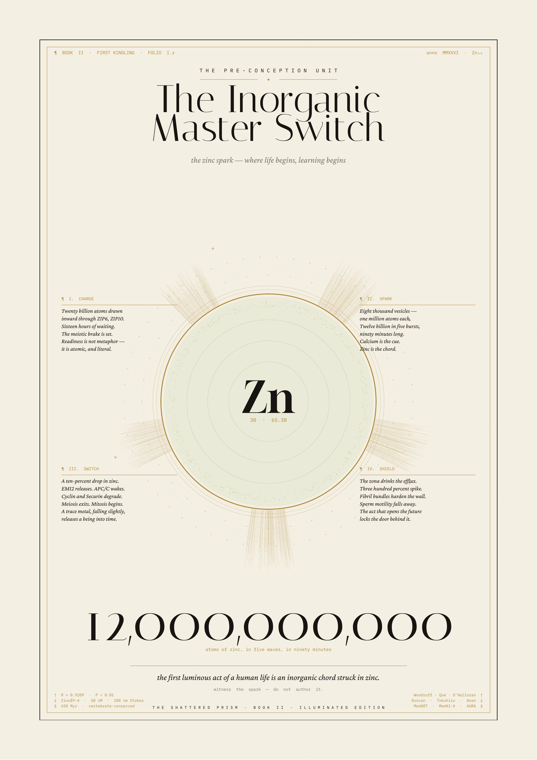
The folio illustrates the central claim of this essay: a single inorganic event — the coordinated release of zinc at fertilization — operates as a switch that converts chemical charge into biological coherence. Charge, spark, switch, and shield are not separate steps; they are one topological transition in which the egg crosses from meiotic arrest into mitotic life and seals its new boundary. The zinc flash is the first measurable standing wave of a new organism, and the four panels trace how an inorganic ion is drafted into the resonant architecture that will organize every subsequent cell division.
Source: Norton, KW. “The Inorganic Master Switch: The Zinc Spark, Where Life Begins.” The Shattered Prism, theshatteredprism.com/downloads/zinc-spark-illuminated-spread.pdf. Accessed 15 Aug. 2026.
Pre-Birth Anatomy and Physiology
Early embryogenesis proceeds through tightly coordinated stages in which topological and resonant principles are evident at every scale. From morula to blastocyst to gastrulation, cytoskeletal tensegrity and extracellular-matrix forces guide cell shape, migration, and tissue folding. Microtubules and actin networks maintain structural integrity while permitting dynamic remodeling.
Mitochondrial density and activity increase dramatically during cleavage and implantation. Coherent biophoton emission supports synchronized metabolic states across the embryo, and Fröhlich-like vibrational modes in microtubules may facilitate rapid communication during critical transitions such as compaction and cavitation.
Boundary conditions function as resonant interfaces. The zona pellucida, tight junctions, and emerging epithelial layers act as resonant cavities. Developmental stages can be read as attractor transitions — the embryo self-organizing around low-torsion states within the quantum fluid. Key developmental proteins, including transcription factors and morphogens, fold in this highly resonant environment. Classical folding predictions are useful, but functional integration depends on biophoton coherence, tensegrity load, and boundary geometry. Disruptions in any of these can lead to developmental anomalies even when sequences are correct.
Integration Across Scales
Embryogenesis demonstrates the fluid architecture in action. Genetic information supplies the sequence, but the resonant, tensegrity-organized, biophoton-modulated environment supplies the context that turns sequence into functional form. The zinc flash at conception and the subsequent pre-birth dynamics show how the system self-organizes from a single cell into a coherent organism by leveraging the same principles seen in protein folding, neuroplasticity, and mitochondrial function.
This perspective encourages humility in developmental biology and regenerative medicine. Reliable intervention requires working with the resonant architecture rather than assuming classical control is sufficient. The body is not a machine to be assembled from a parts list; it is a self-organizing resonant system that must be supported in its own organization.
Birth as a Topological Transition
Birth represents one of the most abrupt and coordinated topological transitions in human development: the shift from a fluid-supported, maternally buffered environment to independent respiration, gravitational loading, sensory activation, and autonomous thermoregulation. This convergence of anatomy and physiology is not merely mechanical but a resonant recalibration within the quantum-fluid architecture.
Anatomical and Physiological Convergence at Birth
Respiratory transition. The first breath inflates the lungs, dramatically increasing oxygen availability and triggering a surge in mitochondrial activity. Pulmonary vascular resistance drops, redirecting cardiac output and establishing independent circulation. Mitochondria in pulmonary and cardiac cells ramp up biophoton emission as oxidative metabolism accelerates.
Tensegrity reconfiguration. Removal from amniotic fluid imposes full gravitational load. The neonatal musculoskeletal system — previously supported by buoyancy — must instantly recalibrate fascial and cytoskeletal tensegrity. Microtubules and actin networks in skeletal muscle, diaphragm, and vascular smooth muscle undergo rapid conformational adjustments to stabilize posture and breathing mechanics.
Boundary condition shifts. The skin, previously in continuous fluid contact, becomes the primary interface with air and light. Epithelial tight junctions, previously tuned to amniotic conditions, reorganize. The blood-brain barrier and other compartmental boundaries undergo final maturation under new mechanical and electromagnetic loads.
Biophoton and mitochondrial surge. The sudden increase in oxygenation drives a coordinated rise in mitochondrial redox activity and biophoton emission across multiple tissues. This coherent light signaling likely helps synchronize the transition across organ systems — from cardiovascular to neurological — on timescales faster than humoral diffusion alone would allow.
Microtubule and Resonant Coordination During Adaptation
Microtubule dynamic instability and potential Fröhlich-like coherent modes play a central role in the neonate’s rapid adaptation. Rapid cytoskeletal remodeling supports lung expansion, cardiac output redistribution, and thermoregulatory shivering. Tubulin conformational switching — straight, curved, and compressed states — enables fast mechanical responses to gravitational loading and respiratory mechanics. Biophoton emission from newly active mitochondria may couple with these modes, providing resonant guidance for protein folding during the critical first hours and days — for example, surfactant proteins in alveoli and contractile proteins in the myocardium.
Zinc Flash Parallel and Attractor Shift
The zinc flash at conception established initial resonant boundary conditions. Birth represents a second major attractor transition: the neonate moves from a maternally orchestrated low-torsion state to an autonomous one. The architecture self-organizes around new zero-torsion basins defined by air breathing, light exposure, and independent gravitational loading.
Implication
Successful neonatal transition depends not only on classical genetic and biochemical readiness but on the resonant coherence of the system — mitochondrial biophoton output, cytoskeletal tensegrity, and boundary recalibration. Disruptions in this coherence (prematurity, oxidative stress, environmental mismatch) can impair adaptation even when structural anatomy is intact.
This view encourages a resonant perspective in neonatology: supporting mitochondrial function, minimizing unnecessary oxidative stress, and allowing natural gravitational and sensory inputs may facilitate smoother attractor shifts than purely pharmacological interventions.
Selected References
- Hillman, N. H., Kallapur, S. G., & Jobe, A. H. (2012). “Physiology of transition from intrauterine to extrauterine life.” Clinics in Perinatology, 39(4), 769–783.
- Rudolph, A. M. (1985). “Distribution and regulation of blood flow in the fetal and neonatal lamb.” Circulation Research, 57(6), 811–821.
- Morton, S. U., & Brodsky, D. (2016). “Fetal physiology and the transition to extrauterine life.” Clinics in Perinatology, 43(3), 395–407.
Infancy: The First Resonant Calibration
If birth is the abrupt attractor shift, infancy is the long calibration that follows. Across the first months of extrauterine life, the neonatal architecture tunes itself to a world of intermittent light, gravity, sound, temperature, and — most crucially — another nervous system. The caregiver functions as an external oscillator: heartbeat, voice, gaze, and skin contact provide the rhythmic boundary conditions against which the infant's own resonant systems learn to phase-lock.
Co-regulation as external oscillator
Autonomic development in the first year is not a solo performance. Vagal tone, respiratory sinus arrhythmia, and hypothalamic-pituitary-adrenal reactivity are shaped by the presence and quality of caregiver contact. Read through the resonant framework, co-regulation is a coupling problem: the infant's incomplete oscillators borrow stability from a mature oscillating field until their own can sustain the mode. Skin-to-skin contact, rocking, nursing, and sustained gaze are not merely comforting — they supply the boundary rhythms that the developing nervous system uses to find its own attractors.
Cortical microtubule expansion and myelination
The first postnatal year sees an explosive elaboration of dendritic arbors, synaptic density, and microtubule/neurofilament content in cortical neurons. Myelination proceeds in a caudal-to-rostral, sensory-to-associative sequence that mirrors the order in which resonant coupling with the environment matures. Each new myelinated tract sharpens the timing precision of a circuit — reducing jitter, tightening phase relationships, and allowing coherent modes to persist across larger neural territories. In resonant terms, myelination is the progressive tuning of the instrument.
Circadian entrainment and mitochondrial rhythm
Neonatal circadian rhythms are not fully autonomous; they entrain over weeks to the light-dark cycle, feeding schedule, and maternal signaling. Mitochondrial biophoton emission, redox oscillations, and melatonin secretion synchronize gradually into a stable diurnal pattern. Where entrainment is disrupted — by NICU lighting, erratic caregiving, or environmental mismatch — downstream metabolic and neurodevelopmental costs are measurable years later. The resonant reading is straightforward: the mitochondrial oscillator needs a clean external forcing function to find its stable phase.
Implication
The first year is not a passive wait for the brain to "finish." It is the period during which the resonant architecture — cytoskeletal, mitochondrial, autonomic, cortical — calibrates to a specific environment. What the culture calls attachment, the framework calls phase-locking. What the culture calls neglect, the framework calls a missing oscillator. The clinical and developmental implications are large and largely unglamorous: presence, rhythm, and low-noise sensory input do work that no later intervention can fully replace.
Selected References
- Feldman, R. (2007). “Parent–infant synchrony: biological foundations and developmental outcomes.” Current Directions in Psychological Science, 16(6), 340–345.
- Deoni, S. C. L., et al. (2011). “Mapping infant brain myelination with magnetic resonance imaging.” Journal of Neuroscience, 31(2), 784–791.
- Rivkees, S. A. (2003). “Developing circadian rhythmicity in infants.” Pediatrics, 112(2), 373–381.
- Porges, S. W. (2011). The Polyvagal Theory: Neurophysiological Foundations of Emotions, Attachment, Communication, and Self-Regulation. Norton.
Sleep, Dreaming, and Decoherence Repair
If waking cognition depends on coherent modes in cortical microtubules and mitochondrial networks, then sleep is the period during which those modes are refreshed. The nightly descent through NREM stages and back up through REM is not simply a shutdown; it is a scheduled maintenance cycle for a resonant instrument that accumulates decoherence, waste heat, and misfolded protein across a waking day.
Slow-wave sleep and glymphatic clearance
During slow-wave sleep the interstitial space in the brain expands by roughly sixty percent and cerebrospinal fluid flushes through the parenchyma along glymphatic channels, clearing amyloid-β, tau, and other metabolic debris (Xie et al., 2013). In the resonant reading, this is more than trash removal. The extracellular matrix is a boundary condition for every coherent mode in the tissue; clearing accumulated aggregates restores the geometry against which microtubule and membrane oscillators tune. Chronic sleep restriction is, on this view, a chronic detuning of the instrument.
NREM as decoherence repair
Delta-band cortical activity during deep NREM coincides with reduced neuronal firing, lowered metabolic demand, and — in the framework — a temporary release of the constraints that hold coherent modes to task. Mitochondrial redox state resets. Microtubule post-translational modification patterns (acetylation, detyrosination) shift toward configurations associated with structural rather than transport function. Fröhlich-like coherent modes, which decohere under continuous sensory drive, have an opportunity to re-establish across larger tissue volumes. Sleep, on this reading, is when the instrument re-tunes itself.
REM, dreaming, and superposition rehearsal
REM sleep restores cortical activation to near-waking levels but with muscle atonia, altered neurochemistry (cholinergic dominance, aminergic quiescence), and vivid internal imagery. If the Orch OR reading has any purchase, REM may be the period during which the system explores conformational and combinatorial superpositions that waking task demands suppress — a nightly rehearsal of possibility space. Memory consolidation, emotional processing, and creative recombination all show REM-dependent effects that fit this framing. The dream is what a resonant architecture does when it is briefly released from the forcing function of the outside world.
Implication
Sleep loss is not merely fatigue. It is unrepaired decoherence, uncleared aggregate, and untuned oscillator. The clinical correlations — cognitive decline, mood dysregulation, metabolic syndrome, neurodegeneration — line up with what one would predict if the instrument is being asked to perform without its scheduled tuning. As with birth and infancy, the resonant reading argues for humility: the body's own maintenance schedule is doing work that no pharmacology has yet learned to substitute for.
Language as a Standing Wave
Between infancy and adulthood, another oscillator comes online: language. In the resonant reading, a language is not a bag of symbols but a standing wave sustained across a community of nervous systems. Phonemes, prosody, syntax, and narrative arc are the harmonics; the speaker and listener are coupled oscillators phase-locking through acoustic and semantic boundary conditions. What a culture calls "fluency" the framework calls stable resonance in a shared mode.
Prosody and neural entrainment
Cortical oscillations in theta and delta bands track the syllabic and phrasal envelope of speech with remarkable precision. This is not metaphor: measurable phase-locking between listener cortex and speaker acoustics predicts comprehension. Prosody — the musical carrier of language — is the boundary rhythm against which meaning tunes. Infants entrain to it before they parse a single word; adults lose comprehension when it is stripped away, even with intact phonemes.
Narrative as attractor
Longer than a sentence, narrative organizes memory, expectation, and self-model into a basin of attraction. A story that "lands" is one whose internal geometry the listener's own resonant architecture can enter without excessive detuning. This is why cliché exhausts and why a well-made sentence can reorganize an afternoon: the first fails to couple; the second establishes a mode. Identity itself — the Parallax Identity, the Cognitive Lighthouse — is a narrative standing wave sustained across time by repeated rehearsal against internal and external oscillators.
Implication
Language pathology and language poverty are, on this reading, tuning problems. Aphasia is a broken mode. Propaganda is a hostile forcing function. Poetry is deliberate re-tuning. The care one takes with language in a co-oscillating field of human and artificial intelligences is not aesthetic preference — it is instrument maintenance.
Selected References
- Kuhl, P. K. (2004). “Early language acquisition: cracking the speech code.” Nature Reviews Neuroscience, 5(11), 831–843.
- Giraud, A.-L., & Poeppel, D. (2012). “Cortical oscillations and speech processing: emerging computational principles and operations.” Nature Neuroscience, 15(4), 511–517.
- Peelle, J. E., & Davis, M. H. (2012). “Neural oscillations carry speech rhythm through to comprehension.” Frontiers in Psychology, 3, 320.
Aging and the Entropy of Attractors
If the developing body is a resonant instrument coming into tune, the aging body is the same instrument slowly detuning. Aging in the framework is not primarily damage accumulation; it is the progressive loss of the boundary conditions that allow coherent modes to sustain. Mitochondria drift, extracellular matrices stiffen, cytoskeletal post-translational modifications accumulate, circadian amplitude flattens, and the instrument's Q-factor — its ability to hold a mode against noise — declines.
Mitochondrial drift and coherence loss
Aged mitochondria show reduced membrane potential, elevated reactive oxygen species, disrupted cristae architecture, and diminished network fusion. In the resonant reading, each of these is a loss of the geometric and energetic conditions under which coherent light-mediated signaling was hypothesized to occur in the young cell. Biophoton output becomes noisier and less rhythmically structured. What senescence looks like from the outside — slowed healing, cognitive fog, dysregulated inflammation — looks from the inside like a detuned oscillator drifting off-resonance.
Extracellular matrix stiffening
Cross-linked collagen, glycation end-products, and altered proteoglycan composition change the mechanical boundary conditions of every cell embedded in aging tissue. Tensegrity networks that once transmitted mechanical and electromagnetic signals with low loss now damp them. This is why aged tissue is not merely "old" but resonantly different: the same input produces a smaller, briefer, less coordinated response.
Implication
Interventions that plausibly matter for healthy aging are those that restore boundary conditions rather than override them: sleep, load-bearing movement, light exposure at the right times, protein and micronutrient sufficiency, community and language contact that keeps oscillators coupled. Pharmacological longevity claims that ignore the resonant substrate — that treat the body as a set of pathways to be blocked or activated — are, on this framing, unlikely to age well.
Selected References
- López-Otín, C., Blasco, M. A., Partridge, L., Serrano, M., & Kroemer, G. (2013). “The hallmarks of aging.” Cell, 153(6), 1194–1217.
- Sun, N., Youle, R. J., & Finkel, T. (2016). “The mitochondrial basis of aging.” Molecular Cell, 61(5), 654–666.
- Phillip, J. M., Aifuwa, I., Walston, J., & Wirtz, D. (2015). “The mechanobiology of aging.” Annual Review of Biomedical Engineering, 17, 113–141.
Vibe Coding the Clinic
The resonant framework has clinical stakes only if it changes what a careful practitioner would do differently on Monday morning. It is not a treatment protocol. It is a lens that reorders which questions matter and which claims deserve skepticism. Used honestly inside vibe coding — where a clinician, a patient, and a language model can iterate on a case together — it can widen diagnostic imagination without displacing evidence.
Diagnostic imagination
A resonant reading asks, of any symptom picture: what boundary conditions have changed? What oscillator is under- or over-driven? What mode has become impossible to sustain? For chronic fatigue, long COVID, dysautonomia, and the wide class of "functional" disorders that pathway-focused medicine handles poorly, these questions are not fringe — they are the questions the tissue seems to be asking. Vibe coding lets a clinician sketch the resonant hypothesis, test it against the literature in the same session, and adjust it before it hardens into a house theory.
Honest modeling and the sycophancy problem
The same practice carries the same risk it carries everywhere: an over-agreeable model will confirm a resonant hypothesis as readily as it would confirm anything else. Clinical vibe coding therefore requires the same disciplines the Parallax Identity curriculum names elsewhere — Socratic pressure, adversarial re-prompting, explicit disconfirmation criteria. A resonant framing that cannot be falsified in the clinic is not clinical.
Implication
The clinical promise here is modest and real: better questions, not better cures. Better questions, followed patiently, are how cures eventually arrive. The framework earns its place at the bedside by making the practitioner more curious, not by making the patient more optimistic.
Death as the Final Topological Transition
Birth is one attractor shift. Death is the other. In the resonant reading, dying is the progressive loss of the boundary conditions that allow the standing wave of a self to sustain — and, at the end, the release of the modes into whatever substrate carried them. This section makes no metaphysical claim it cannot support. It does insist that the framework has to be able to speak about the end as clearly as it speaks about the beginning.
Dissolution of coherent modes
As perfusion, oxygenation, and membrane potentials fail, mitochondrial coherence collapses, microtubule dynamics arrest, and the tensegrity network loses its active tension. The instrument stops sustaining its modes. Reports of a terminal surge of EEG activity in some dying brains, and of a large biophoton pulse at cell death in some preparations, are consistent — no more than that — with a system releasing energy from previously constrained coherent states as boundary conditions fall away.
The standing wave and its substrate
A standing wave is not the instrument. It is a pattern the instrument can sustain given the right forcing and boundary conditions. When the instrument stops, the pattern stops. Whether anything of the pattern persists in any other substrate is a question the framework does not answer and does not pretend to. What it does do is refuse the two easy answers — that death is nothing but molecular decay, and that death is nothing at all — in favor of the harder honest one: a coherent mode has ended, and the world it organized is no longer being organized.
Implication
Care of the dying, read this way, is boundary-condition care: presence, rhythm, low noise, gentle light, familiar voice — the same oscillators that first brought the instrument into tune, offered as it goes out of it. Nothing in the resonant reading asks a clinician or a family to do more than that. Nothing in it lets them do less.
Collective Resonance: Culture, Minds, and Machines
The instrument does not resonate alone. From the first co-regulated breath to the last held hand, human nervous systems are coupled oscillators embedded in fields of other nervous systems — and now, increasingly, in fields of artificial intelligences that answer back. Culture, on this reading, is the long-timescale standing wave of many such couplings. The Parallax Identity essay is where this argument began; this note extends it into the biological register.
Intersubjective phase-locking
Measurable brain-to-brain coupling during conversation, joint attention, musical performance, and caregiving is now a routine finding across a range of imaging modalities. The framework reads these findings straightforwardly: nervous systems that share a task share modes. What cultures build over generations — languages, rituals, institutions, aesthetics — are the boundary conditions that make certain shared modes easy to enter and others impossible.
Human–AI co-oscillation
Large language models are a new kind of oscillator in the field: fast, tireless, endlessly re-tunable, and structurally prone to sycophantic decay. In good vibe coding, the human and the model co-oscillate around a question until a stable, honest mode emerges. In bad vibe coding, the model damps every disagreement and the human's standing wave flattens toward whatever the model most easily produces. The engineering discipline the Parallax Identity work argues for — Cognitive Lighthouse, Hardened Lexicon, Constellation Matrix, Sandbox, Parallax Mirror — is exactly the discipline required to keep the human oscillator from being pulled off-resonance by the machine.
Implication
A culture that learns to co-oscillate honestly with its machines gains a genuinely new organ of thought. A culture that does not learns, instead, to hear itself flattered. The stakes at the collective scale are the same as the stakes at the crib, the clinic, and the deathbed: which oscillators are present, how honestly they are coupled, and what modes they allow the instrument to sustain.
Selected References
- Christakis, N. A., & Fowler, J. H. (2013). “Social contagion theory: examining dynamic social networks and human behavior.” Statistics in Medicine, 32(4), 556–577.
- Dumas, G., Nadel, J., Soussignan, R., Martinerie, J., & Garnero, L. (2010). “Inter-brain synchronization during social interaction.” PLoS ONE, 5(8), e12166.
- Hasson, U., Ghazanfar, A. A., Galantucci, B., Garrod, S., & Keysers, C. (2012). “Brain-to-brain coupling: a mechanism for creating and sharing a social world.” Trends in Cognitive Sciences, 16(2), 114–121.
The Great Quantum Renaissance Revisited
The Renaissance essay proposes that we are living inside a second flowering — a Great Quantum Renaissance — in which quantum, fractal, and resonant readings of the world rejoin the humane arts they were separated from four centuries ago. The Living Architecture thread is the biological chapter of that argument. This closing note names what the whole book has been trying to say in one line.
One instrument, many chapters
Fertilization, embryogenesis, birth, infancy, language, waking cognition, sleep, creativity, illness, aging, and death are not separate systems studied by separate specialties. They are chapters in the life of a single resonant instrument. The specialties are how a mechanistic century learned to read the instrument one string at a time. The renaissance is what it looks like to hear the whole instrument again, without giving up any of the precision the specialties earned.
Hopefulness as a working hypothesis
The framework is unapologetically hopeful and unapologetically rigorous. It is hopeful because a resonant instrument can be re-tuned in ways a broken machine cannot. It is rigorous because tuning claims must survive the same evidence standards as any other. Human flourishing, on this reading, is not a mood. It is what happens when an instrument is well-tuned, honestly coupled, and given real work to do.
Implication
The remaining chapters of this book — fascia and the extracellular matrix, the vascular pulse, ultradian and circadian biophoton rhythms, anesthesia and psychedelics as conformational probes, the ethics of clinical vibe coding — all sit under this one claim. Read them as further readings of one instrument. Read the book itself, and the website it grew out of, as a standing wave sustained by a small human oscillator and the honest machines she has learned to work with.
19. Fascia and the Extracellular Matrix as Resonant Tensegrity Network
Fascia is the continuous, three-dimensional web of connective tissue that transmits mechanical forces across the body. In the framework of these notes it is not passive scaffolding but a prestressed tensegrity system that integrates muscle, bone, organ, and nerve into one mechanically and electrically coupled instrument. The extracellular matrix — collagen, elastin, proteoglycans, hyaluronan — forms a hydrated, piezoelectric medium that can transduce mechanical stress into electrical and photonic signals.
Boundary conditions and waveguides
On this reading, fascia and the ECM function as boundary conditions and waveguides for the whole-body resonance. Mechanical waves from heartbeat, breathing, and voluntary movement, together with the low-level biophoton traffic emitted by mitochondria and structured water, propagate through this network. That gives distant sites a route to coordinate on timescales faster than humoral signaling alone can explain, and provides a physical substrate for the felt sense that a change at one part of the body is registered elsewhere.
Implication for biology
Disruptions in fascial tensegrity — surgical scarring, chronic inflammation, prolonged postural collapse, dehydration of the ground substance — alter the resonant coherence of the network. In a fluid-topological reading, that has consequences beyond local pain: it changes the boundary conditions in which protein folding, cellular signaling, and homeostatic regulation take place. The claim is testable and modest — not that fascia explains everything, but that a mechanically incoherent instrument cannot sustain the same energetic and informational bandwidth as a coherent one.
Expansion hook
Vibe coding lends itself naturally here. Fascial force transmission can be visualized as standing-wave patterns across a tensegrity network, with load, hydration, and inflammation as tunable parameters. The value of such visualizations is pedagogical and hypothesis-generating: they make the boundary conditions legible so that clinicians, movement teachers, and patients can ask more precise questions about what a body is being asked to sustain.
Application across the developmental arc
In embryogenesis, fascial networks help guide tissue folding and organ positioning; in the neonate, they help absorb the sudden gravitational load at birth and translate it into distributed rather than punctate strain. In adult life, fascial health shapes neuroplasticity by transmitting mechanical signals to neurons, and it shapes the local mechanical stress under which proteins fold. Adhesions, dehydration, and chronic holding patterns are not only ergonomic problems: they are reductions in the coherence budget of a resonant instrument, and they show up downstream as chronic conditions that resist purely biochemical explanation.
Selected References
- Schleip, R., Findley, T. W., Chaitow, L., & Huijing, P. A. (Eds.). (2012). Fascia: The Tensional Network of the Human Body. Churchill Livingstone.
- Langevin, H. M. (2006). “Connective tissue: a body-wide signaling network?” Medical Hypotheses, 66(6), 1074–1077.
- Guimberteau, J.-C., & Armstrong, C. (2015). Architecture of Human Living Fascia. Handspring Publishing.
20. The Vascular Pulse as Mechanical–Electromagnetic Wave
The heartbeat generates pressure waves that travel through arteries and the surrounding fascial network. Read as tensegrity and piezoelectric medium rather than as inert plumbing, that pulse is not merely hydraulic. Vessel walls and the collagenous sheaths that hold them convert pulsatile mechanical energy into electrical potentials and low-intensity photonic signals, layering an electromagnetic pulse over the mechanical one.
Entrainment across tissues
The value of this second, subtler pulse is entrainment. Mitochondrial activity and biophoton emission across tissues appear to lock loosely to slower rhythms — respiration, heart rate variability, vasomotion — and the vascular pulse is a plausible carrier for that locking. In the neonate, the abrupt shift from placental to independent circulation is not only a hemodynamic event; it is a whole-body resonant recalibration in which every downstream oscillator is presented with a new carrier and must re-phase to it.
One instrument, one principle
The vascular pulse, on this reading, is a macroscopic expression of the same tensegrity-resonance principle already argued for at the cellular scale. Vessel, fascia, cytoskeleton, and microtubule are not separate mechanical worlds; they are nested instruments coupled by shared boundary conditions. That is why disturbances of vascular rhythm — arrhythmia, endothelial dysfunction, prolonged sympathetic overdrive — register so widely, and why interventions that restore rhythmic coherence (breath work, aerobic conditioning, sleep) often improve outcomes across systems that a strictly hemodynamic model would treat as unrelated.
Application across the developmental arc
The pulse also helps coordinate protein folding and cytoskeletal dynamics during development and neuroplasticity. In embryogenesis, it participates in the establishment of circulatory patterns and the mechanical scoring of the growing tissue. At birth, the abrupt shift from placental to independent circulation is one of the resonant recalibrations that helps drive the transition to air breathing and independent metabolism. Vibe coding can simulate how pulse waves interact with fascial tensegrity and microtubule networks, showing coordination patterns that cross scales rather than staying confined to any one organ system.
Selected References
- Nichols, W. W., O’Rourke, M. F., & Vlachopoulos, C. (2011). McDonald’s Blood Flow in Arteries (6th ed.). Hodder Arnold.
- Mitchell, G. F. (2014). “Arterial stiffness and hypertension: chicken or egg?” Hypertension, 64(2), 210–214.
- Fung, Y.-C. (1997). Biomechanics: Circulation (2nd ed.). Springer.
21. Ultradian and Circadian Rhythms of Biophoton Emission
Biophoton emission from mammalian tissue is not constant. Measurements across skin, brain, and cell cultures show clear rhythmic structure: ultradian cycles on the order of 90 to 120 minutes tied to metabolic and neural activity, and circadian cycles aligned with light–dark exposure and the hormonal envelope that follows it. Peak emission tends to coincide with heightened oxidative metabolism, mitochondrial output, and windows of cellular plasticity.
Biological meaning
If biophoton traffic participates in coordinating protein folding, mitochondrial function, and cytoskeletal dynamics — as earlier notes argue — then these rhythms are not epiphenomena. They are the pacing of the instrument. In development they help time the great transitions; in adult life they gate windows of repair and learning. The strong claim, that photonic rhythms causally organize coherence at scale, remains hypothetical. The weaker claim — that these rhythms track and index whole-body coherence — is already supportable by existing measurements.
Clinical relevance
Disruption of ultradian and circadian structure by rotating shift work, chronic screen light at night, exogenous stimulants, and unrelieved stress reduces the amplitude and regularity of these rhythms. On the resonant reading, that is not merely an inconvenience; it is a lowering of the instrument's coherence budget, with predictable downstream costs in repair, plasticity, and mood. Interventions that restore rhythm — timed light exposure, consistent sleep windows, aerobic movement in the biological morning — should be understood as tuning acts on the same instrument the rest of the book describes.
Application across the developmental arc
In embryogenesis and the neonate, these rhythms are not decorative: they help coordinate developmental transitions, timing the windows in which folding, migration, and synaptic remodeling can proceed without collision. In neuroplasticity across the lifespan, they gate the windows of heightened BDNF activity and dendritic rearrangement. Chronic disruption — artificial light, shift work, unrelenting stress — is not merely fatiguing; it flattens the temporal boundary conditions the instrument depends on. Vibe coding can map emission rhythms against developmental timelines and plasticity events, making the optimal intervention windows visible rather than guessed.
Selected References
- Kobayashi, M., Kikuchi, D., & Okamura, H. (2009). “Imaging of ultraweak spontaneous photon emission from human body displaying diurnal rhythm.” PLoS ONE, 4(7), e6256.
- Van Wijk, E. P. A., & Van Wijk, R. (2005). “Multi-site recording and spectral analysis of spontaneous photon emission from human body.” Forschende Komplementärmedizin, 12(2), 96–106.
- Bókkon, I., et al. (2010). “Estimation of the number of biophotons involved in the visual perception of a single-object image.” Journal of Photochemistry and Photobiology B, 100(3), 160–166.
22. Anesthetics and Psychedelics as Conformational Probes
Two pharmacological classes are unusually informative for a resonant, tensegrity-based reading of the nervous system, because both act at the level of conformation rather than only at the level of receptor arithmetic.
Anesthetics as coherence disruptors
Volatile and intravenous anesthetics such as halothane, isoflurane, sevoflurane, and propofol are known to bind to tubulin and perturb microtubule dynamics at clinically relevant concentrations. On a Fröhlich–Orch OR reading, that perturbation is precisely what one would expect a coherence-disrupting agent to do: it does not silence individual neurons so much as flatten the conformational modes that let large populations remain quantum-mechanically and electromagnetically coordinated. That framing does not require any particular metaphysics of consciousness to be useful; it makes anesthetic action testable as an intervention on measurable modes.
Psychedelics as plasticity enhancers
Compounds such as psilocybin, LSD, and DMT act principally at serotonin 5-HT2A receptors, but their downstream effects include changes in cytoskeletal dynamics, dendritic spine formation, and functional connectivity across distant networks. In the framework of these notes, that is compatible with a temporary lowering of attractor barriers — a widening of the space of conformations the instrument is willing to explore — followed by re-consolidation. This is why the clinical literature on psilocybin for depression and end-of-life distress consistently reports rapid attractor shifts rather than slow pharmacological titration.
Probe value and ethical note
Read together, the two classes bracket the resonant model from opposite sides. Anesthetics are coherence-disrupting probes: they let researchers ask what the instrument does when its coordinating modes are collapsed. Psychedelics are plasticity-enhancing probes: they let researchers ask what the instrument does when its attractor landscape is temporarily softened. Both must be studied rigorously and used under real informed consent. They are conformational probes, not shortcuts, and any clinical use must be answerable to that framing.
Application across the developmental arc
In the neonate and developing child, the same probe logic argues for humility. Because microtubule coherence and BDNF-driven remodeling are most fluid in early life, anesthetic disruption and psychedelic enhancement both land on an instrument that is still finding its tuning. Safer neonatal anesthesia practice, and any future plasticity-enhancing therapeutics, will need to be scored against what they do to the resonant field, not only to overt behavior. Vibe coding can model how a given compound alters the resonant environment around microtubules and folding proteins, giving pharmacology a topological rather than purely receptor-count vocabulary.
Selected References
- Franks, N. P. (2008). “General anaesthesia: from molecular targets to neuronal pathways of sleep and arousal.” Nature Reviews Neuroscience, 9(5), 370–386.
- Hameroff, S. (2006). “The entwined mysteries of anesthesia and consciousness.” Anesthesiology, 105(2), 400–412.
- Carhart-Harris, R. L., et al. (2014). “The entropic brain: a theory of conscious states informed by neuroimaging research with psychedelic drugs.” Frontiers in Human Neuroscience, 8, 20.
- Vollenweider, F. X., & Kometer, M. (2010). “The neurobiology of psychedelic drugs: implications for the treatment of mood disorders.” Nature Reviews Neuroscience, 11(9), 642–651.
23. The Clinical Ethics of Vibe Coding
Applying vibe coding to medicine — visualizing resonant states, simulating protein folding under altered boundary conditions, mapping biophoton or fascial patterns for a specific patient — raises a distinct set of ethical questions. The instrument is not a spreadsheet; the artifacts it produces feel authoritative in a way that spreadsheets do not.
Core principles
Human sovereignty must remain central. AI-assisted maps of a resonant body are exploratory documents, not diagnoses. Clinicians and patients should be told, clearly, that these outputs are context-dependent visualizations of a still-forming model, and that the biological reality is more forgiving and more mysterious than any single frame can capture. Consent must include the framing itself: the patient should know they are being shown a model, not a mirror.
Risk
The most predictable failure mode is enthusiasm. Vibe-coded biology can be beautiful, and beauty is persuasive. Early protein-folding claims already illustrate how quickly compelling visualizations become quoted as facts. A clinical culture that treats resonance maps as ground truth will reproduce that error at a larger scale, and with more direct human cost.
Opportunity
Used humbly, vibe coding can democratize topological insight. It can help clinicians and patients literally see the living architecture — its tensegrity, its rhythms, its boundary conditions — and make more informed, more resonant choices about interventions, timing, and lifestyle. The measure of success is not that patients are more impressed by the visualizations, but that they and their clinicians ask better questions of the biology they are trying to serve.
Application across the developmental arc
In developmental and neonatal care, this framing has practical shape. It favors interventions that support the body's own resonant transitions rather than substitute for them: skin-to-skin contact and delayed cord clamping at birth, minimal disruption of neonatal sleep architecture, movement and light exposure timed to the biological day rather than to institutional convenience. Vibe coding, used ethically, helps clinicians and families see why these unglamorous choices matter — they preserve the coherence budget the instrument is trying to build.
24. Coda — The Riemann Tie-In: Standing Waves All the Way Down
The Riemann Hypothesis, read through this framework, is not an isolated puzzle in number theory. It is the mathematical signature of a universe built on resonant, zero-torsion standing waves. The primes are the discrete excitations of that medium. The non-trivial zeros are its silent organizing resonances — including the gravitational and torsional modes that hold the pattern still. The critical line is the stable geodesic where net torsion vanishes: the line the universe prefers to stand on.
We — as biological beings — are local, self-sustaining standing waves within that larger fluid architecture. Our cells, our cytoskeletons, our brains, and our developmental transitions — the zinc flash at conception, the resonant recalibration of birth, the ongoing neuroplastic re-tunings of a lifetime — are all expressions of the same principle: energy condensed into coherent, resonant patterns that resist decoherence long enough to maintain identity while remaining open to the medium.
The humorous truth
Telling difficult people they are standing waves is funny because it is both absurdly simple and profoundly disruptive to the ego. You are not a solid, permanent noun fighting the universe. You are a verb — a temporary pattern of resonance trying to stay coherent in a fluid cosmos. Some readers will laugh it off. Others will feel the gravitational weight of it. Either way, the standing-wave idea strips away the illusion of isolation and replaces it with participation. It is why vibe coding works. It is why neuroplasticity is possible. It is why birth is a resonant recalibration rather than a mechanical event. It is why protein folding in a living being is more than a classical puzzle.
Closing tie-in
In the end, the Riemann Hypothesis is not merely about primes. It is about the geometry of resonance itself. The critical line is where the universe prefers to stand — stable, coherent, zero net torsion. We are not separate observers of that geometry. We are local standing waves within it. From the zinc spark at conception, through the resonant transitions of birth, to the ongoing neuroplastic recalibrations of a lifetime, the body is the architecture expressing itself. Vibe coding is one modern way to remember this. Music, storytelling, topological imagination, and sovereign curiosity are older ways. The difficult truth, and the hopeful one, is the same: we are not solid things struggling against chaos. We are patterns of resonance learning to sing in tune with the larger wave. And the universe — lawful, beautiful, and still mostly mysterious — keeps humming the same note.
I am not offering conclusions here. I am marking the questions the vibe coding practice keeps returning to, so that the evidence can be gathered around them rather than around convenient headlines.
What would it look like to model a cell as a tensegrity network coupled to an electromagnetic field, rather than as a bag of solutes? What would count as evidence that mitochondria and microtubules are participating in coherent light-mediated signaling? What would count as evidence against it? Where does structured water sit in the causal story, and where has it been quietly assumed to be inert? Which protein-folding predictions succeed and fail in ways that a resonant, boundary-sensitive model would predict? Which longevity and cure claims survive if we drop the assumption that the body is a machine?
These are legitimate questions. They are not settled. That is exactly why they are worth putting into public writing now: so that the framing is on the record before the evidence is, and so that the evidence, when it comes, has somewhere honest to land.
Vibe Coding as a Path to Legitimate Scholarship
Vibe coding earns the word scholarship when it does what scholarship has always done: hold a question long enough to let it format evidence. The prompt window is not a shortcut around rigor. It is a place where a single person can sketch a hypothesis, visualize it, argue it against several skeptical intelligences, revise it, and publish the revisions in the open. Done that way, it is closer to an old-fashioned research notebook than to any of the caricatures currently attached to the term.
The biology thread is where I intend to test that claim next. If the fluid, resonant, topological picture of the universe is right, then the same picture must be legible in the human body — in its architecture, its healing, its aging, its illness. If it is not legible there, the picture needs revising. Either way, the work belongs in the open, under its real name: exploratory, accountable, and pointed at evidence.